


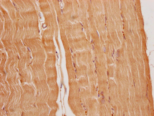
Immunohistochemical staining of human skeletal muscle shows moderate cytoplasmic positivity in myocytes.
Immunohistochemical staining of human skeletal muscle shows moderate cytoplasmic positivity in myocytes.
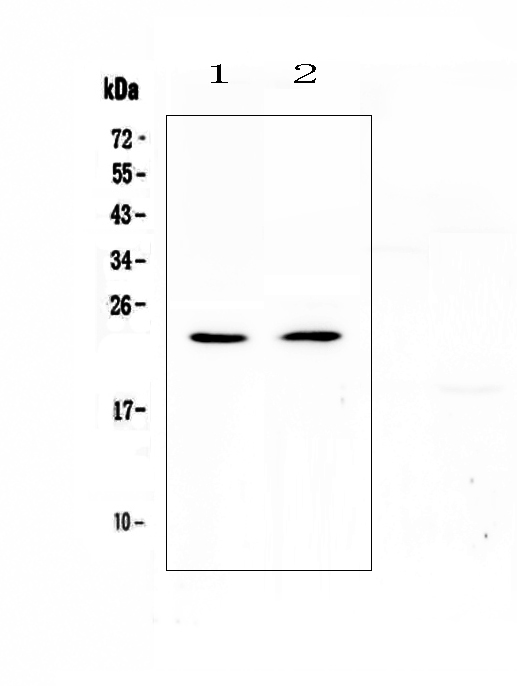
Anti-FNDC5 Antibody
HPA051290
ApplicationsImmunoHistoChemistry
Product group Antibodies
ReactivityHuman
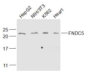
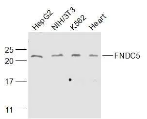
TargetFNDC5
Overview
- SupplierAtlas Antibodies
- Product NameAnti-FNDC5 Antibody
- Delivery Days Customer4
- ApplicationsImmunoHistoChemistry
- CertificationResearch Use Only
- ClonalityPolyclonal
- ConjugateUnconjugated
- Gene ID252995
- Target nameFNDC5
- Target descriptionfibronectin type III domain containing 5
- Target synonymsFRCP2, irisin, fibronectin type III domain-containing protein 5, fibronectin type III repeat-containing protein 2
- HostRabbit
- IsotypeIgG
- Protein IDQ8NAU1
- Protein NameFibronectin type III domain-containing protein 5
- Scientific DescriptionRecombinant Protein Epitope Signature Tag (PrEST) antigen sequence
- ReactivityHuman
- Storage Instruction-20°C,2°C to 8°C
- UNSPSC41116161
Datasheet
MSDS
Related products
Product group Antibodies
anti-Irisin, pAb (IN102)AG-25B-0027
ApplicationsWestern Blot
ReactivityHuman, Monkey, Mouse, Rat
TargetFNDC5
- SizePrice
Product group Antibodies
Anti-FNDC5 Antibody Picoband(r)A02538-1-CARRIER-FREE
ApplicationsWestern Blot, ELISA
ReactivityHuman, Mouse, Rat
TargetFNDC5
- SizePrice
Product group Antibodies
Anti-FNDC5 (-C) (94-111) Antibody130-10570
ApplicationsELISA
ReactivityHuman
TargetFNDC5
- SizePrice
Product group Antibodies

ApplicationsImmunoHistoChemistry, ImmunoHistoChemistry Paraffin
ReactivityHuman, Mouse, Rat
TargetFNDC5
- SizePrice
Product group Antibodies
References
FNDC5 Polyclonal AntibodyBS-8486R
ApplicationsImmunoFluorescence, Western Blot, ELISA, ImmunoCytoChemistry, ImmunoHistoChemistry, ImmunoHistoChemistry Frozen, ImmunoHistoChemistry Paraffin
ReactivityCanine, Chicken, Human, Mouse, Rabbit, Rat
TargetFNDC5
- SizePrice
Product group Antibodies
FNDC5 AntibodyCSB-PA836707OA01HU
ApplicationsImmunoFluorescence, ELISA, ImmunoHistoChemistry
ReactivityHuman
TargetFNDC5
- SizePrice
Product group Antibodies
ApplicationsImmunoPrecipitation, Western Blot, ImmunoCytoChemistry, ImmunoHistoChemistry
ReactivityMouse, Porcine, Rat
TargetFNDC5
- SizePrice
Product group Antibodies
FNDC5 antibodyGTX03463
ApplicationsWestern Blot
ReactivityHuman, Mouse
TargetFNDC5
- SizePrice