Bio-Connect
Immunofluorescent staining of human cell line Hep G2 shows localization to nucleoplasm.
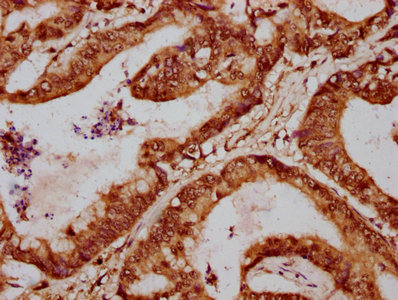
Anti-FOXA3 Antibody

HPA054034
Atlas Antibodies
ApplicationsImmunoCytoChemistry, Chip-exoSeq
Product group Antibodies
TargetFOXA3
Sign in to order and to see your custom pricing.
Large volume orders?
Order with a bulk request

Overview

  • Supplier
    Atlas Antibodies
  • Product Name
    Anti-FOXA3
  • Delivery Days Customer
    4
  • Applications
    ImmunoCytoChemistry, Chip-exoSeq
  • Certification
    Research Use Only
  • Clonality
    Polyclonal
  • Conjugate
    Unconjugated
  • Gene ID3171
  • Target name
    FOXA3
  • Target description
    forkhead box A3
  • Target synonyms
    FKHH3; fork head-related protein FKH H3; forkhead box protein A3; hepatocyte nuclear factor 3-gamma; HNF3G; HNF-3G; HNF-3-gamma; TCF3G; TCF-3G; transcription factor 3G
  • Host
    Rabbit
  • Isotype
    IgG
  • Protein IDP55318
  • Protein Name
    Hepatocyte nuclear factor 3-gamma
  • Scientific Description
    Recombinant Protein Epitope Signature Tag (PrEST) antigen sequence
  • Storage Instruction
    -20°C,2°C to 8°C
  • UNSPSC
    12352203

Related products

IHC image of CSB-PA008796LA01HU diluted at 1:700 and staining in paraffin-embedded human colon cancer performed on a Leica BondTM system. After dewaxing and hydration, antigen retrieval was mediated by high pressure in a citrate buffer (pH 6.0). Section was blocked with 10% normal goat serum 30min at RT. Then primary antibody (1% BSA) was incubated at 4°C overnight. The primary is detected by a biotinylated secondary antibody and visualized using an HRP conjugated SP system.
Product group Antibodies
Cusabio
FOXA3 AntibodyCSB-PA008796LA01HU
ApplicationsELISA, ImmunoHistoChemistry
TargetFOXA3
  • SizePrice
WB analysis of various samples using GTX04941 FOXA3 antibody. Lane 1 : Rat liver tissue lysate (50 microg) Lane 2 : Rat pancreas tissue lysate (50 microg) Lane 3 : Mouse liver tissue lysate (50 microg) Lane 4 : HeLa whole cell lysate (40 microg) Dilution : 0.5 microg/mL
Product group Antibodies
GeneTex
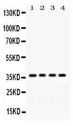
ApplicationsWestern Blot
TargetFOXA3
  • SizePrice
Atlas Antibodies
Protein IDP55318
  • SizePrice
Product group Antibodies
LifeSpan BioSciences
FOXA3 AntibodyLS-C680489
ApplicationsELISA, ImmunoHistoChemistry, ImmunoHistoChemistry Paraffin
TargetFOXA3
  • SizePrice
Figure 1. Western blot analysis of FOXA3 using anti-FOXA3 antibody (PB9805). Electrophoresis was performed on a 5-20% SDS-PAGE gel at 70V (Stacking gel) / 90V (Resolving gel) for 2-3 hours. The sample well of each lane was loaded with 30 ug of sample under reducing conditions. Lane 1: human HepG2 whole cell lysates. After electrophoresis, proteins were transferred to a nitrocellulose membrane at 150 mA for 50-90 minutes. Blocked the membrane with 5% non-fat milk/TBS for 1.5 hour at RT. The membrane was incubated with rabbit anti-FOXA3 antigen affinity purified polyclonal antibody (Catalog # PB9805) at 0.5 microg/mL overnight at 4°C, then washed with TBS-0.1%Tween 3 times with 5 minutes each and probed with a goat anti-rabbit IgG-HRP secondary antibody at a dilution of 1:5000 for 1.5 hour at RT. The signal is developed using an Enhanced Chemiluminescent detection (ECL) kit (Catalog # EK1002) with Tanon 5200 system. A specific band was detected for FOXA3 at approximately 40 kDa. The expected band size for FOXA3 is at 37 kDa.
Product group Antibodies
Boster Bio
ApplicationsFlow Cytometry, ImmunoFluorescence, Western Blot, ImmunoCytoChemistry, ImmunoHistoChemistry
TargetFOXA3
  • SizePrice