

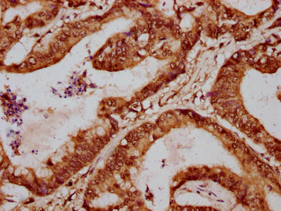
IHC image of CSB-PA008796LA01HU diluted at 1:700 and staining in paraffin-embedded human colon cancer performed on a Leica BondTM system. After dewaxing and hydration, antigen retrieval was mediated by high pressure in a citrate buffer (pH 6.0). Section was blocked with 10% normal goat serum 30min at RT. Then primary antibody (1% BSA) was incubated at 4°C overnight. The primary is detected by a biotinylated secondary antibody and visualized using an HRP conjugated SP system.
IHC image of CSB-PA008796LA01HU diluted at 1:700 and staining in paraffin-embedded human colon cancer performed on a Leica BondTM system. After dewaxing and hydration, antigen retrieval was mediated by high pressure in a citrate buffer (pH 6.0). Section was blocked with 10% normal goat serum 30min at RT. Then primary antibody (1% BSA) was incubated at 4°C overnight. The primary is detected by a biotinylated secondary antibody and visualized using an HRP conjugated SP system.
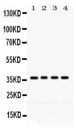
FOXA3 Antibody
CSB-PA008796LA01HU
ApplicationsELISA, ImmunoHistoChemistry
Product group Antibodies
ReactivityHuman
TargetFOXA3
Overview
- SupplierCusabio
- Product NameFOXA3 Antibody
- Delivery Days Customer20
- ApplicationsELISA, ImmunoHistoChemistry
- CertificationResearch Use Only
- ClonalityPolyclonal
- ConjugateUnconjugated
- Gene ID3171
- Target nameFOXA3
- Target descriptionforkhead box A3
- Target synonymsFKHH3, HNF3G, TCF3G, hepatocyte nuclear factor 3-gamma, HNF-3-gamma, HNF-3G, TCF-3G, fork head-related protein FKH H3, forkhead box protein A3, transcription factor 3G
- HostRabbit
- IsotypeIgG
- Protein IDP55318
- Protein NameHepatocyte nuclear factor 3-gamma
- Scientific DescriptionTranscription factor that is thought to act as a pioneer factor opening the compacted chromatin for other proteins through interactions with nucleosomal core histones and thereby replacing linker histones at target enhancer and/or promoter sites (By similarity). Originally described as a transcription activator for a number of liver genes such as AFP, albumin, tyrosine aminotransferase, PEPCK, etc. Interacts with the cis-acting regulatory regions of these genes. Involved in glucose homeostasis; binds to and activates transcription from the G6PC promoter. Binds to the CYP3A4 promoter and activates its transcription in cooperation with CEBPA. Binds to the CYP3A7 promoter together with members of the CTF/NF-I family. Involved in regulation of neuronal-specific transcription. May be involved in regulation of spermatogenesis.
- ReactivityHuman
- Storage Instruction-20°C or -80°C
- UNSPSC41116161
Related products
Product group Antibodies

FOXA3 AntibodyLS-C680489
ApplicationsELISA, ImmunoHistoChemistry, ImmunoHistoChemistry Paraffin
ReactivityHuman
TargetFOXA3
- SizePrice
Product group Antibodies
FOXA3 antibodyGTX04941
ApplicationsWestern Blot
ReactivityHuman, Mouse, Rat
TargetFOXA3
- SizePrice

Product group Antibodies
Anti-FOXA3 AntibodyHPA054034
ApplicationsChIP Chromatin ImmunoPrecipitation, ImmunoCytoChemistry
ReactivityHuman
TargetFOXA3
- SizePrice

Product group Antibodies
Anti-FOXA3 Antibody Picoband(r)PB9805-CARRIER-FREE
ApplicationsFlow Cytometry, ImmunoFluorescence, Western Blot, ImmunoCytoChemistry, ImmunoHistoChemistry
ReactivityHamster, Human, Mouse, Rat
TargetFOXA3
- SizePrice