

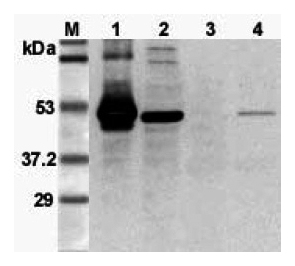
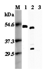
Western blot analysis using anti-FOXP3 (mouse), pAb (Prod. No. AG-25A-0020) at 1:3000 dilution. 1: Mouse FOXP3 (His-tagged). 2: Transfected mouse FOXP3 cell lysate (HEK 293). 3: Mouse T lymphocyte (CD4+) cell lysate. 4: PHA treate
Western blot analysis using anti-FOXP3 (mouse), pAb (Prod. No. AG-25A-0020) at 1:3000 dilution. 1: Mouse FOXP3 (His-tagged). 2: Transfected mouse FOXP3 cell lysate (HEK 293). 3: Mouse T lymphocyte (CD4+) cell lysate. 4: PHA treate
anti-FOXP3 (mouse), pAb
AG-25A-0020
ApplicationsWestern Blot, ELISA
Product group Antibodies
ReactivityMouse
TargetFoxp3
Overview
- SupplierAdipoGen Life Sciences
- Product Nameanti-FOXP3 (mouse), pAb
- Delivery Days Customer10
- ApplicationsWestern Blot, ELISA
- CertificationResearch Use Only
- ClonalityPolyclonal
- Concentration1 mg/ml
- Gene ID20371
- Target nameFoxp3
- Target descriptionforkhead box P3
- Target synonymsJM2, scurfin, sf, forkhead box protein P3, scurfy
- HostRat
- Protein IDQ99JB6
- Protein NameForkhead box protein P3
- Scientific DescriptionFOXP3 is involved in immune system responses. It functions as the master regulator in the development and function of regulatory T cells. FOX proteins belong to the forkhead/winged-helix family of transcriptional regulators and are presumed to exert control via similar DNA binding interactions during transcription. Defects in FOXP3 are the cause of immunodeficiency polyendocrinopathy, enteropathy, X-linked syndrome (IPEX); also known as X-linked autoimmunity-immunodeficiency syndrome. IPEX is characterized by neonatal onset insulin-dependent diabetes mellitus, infections, secretory diarrhea, trombocytopenia, anemia and eczema. - Polyclonal Antibody. Recognizes mouse FOXP3. Detects a band of ~46-49kDa by Western blot. Source: Rat. Applications: ELISA, WB. Liquid. 0.2microm-filtered solution in PBS, pH 7.4. Contains no preservatives. FOXP3 is involved in immune system responses. It functions as the master regulator in the development and function of regulatory T cells. FOX proteins belong to the forkhead/winged-helix family of transcriptional regulators and are presumed to exert control via similar DNA binding interactions during transcription. Defects in FOXP3 are the cause of immunodeficiency polyendocrinopathy, enteropathy, X-linked syndrome (IPEX); also known as X-linked autoimmunity-immunodeficiency syndrome. IPEX is characterized by neonatal onset insulin-dependent diabetes mellitus, infections, secretory diarrhea, trombocytopenia, anemia and eczema.
- ReactivityMouse
- Storage Instruction-20°C,2°C to 8°C
- UNSPSC41116161
Related products

Product group Antibodies
anti-FOXP3 (mouse), mAb (MF333F)AG-20A-0025Y
ApplicationsWestern Blot, ELISA, ImmunoCytoChemistry, ImmunoHistoChemistry
ReactivityMouse
TargetFoxp3
- SizePrice
Product group Antibodies
anti-FOXP3 (mouse), mAb (MF333F)AG-20A-0025Y
ApplicationsWestern Blot, ELISA, ImmunoCytoChemistry, ImmunoHistoChemistry
ReactivityMouse
TargetFoxp3
- SizePrice
Product group Antibodies
anti-FOXP3 (mouse), pAbAG-25A-0035
ApplicationsWestern Blot, ELISA
ReactivityMouse
TargetFoxp3
- SizePrice
Product group Antibodies
anti-FOXP3 (human), mAb (ANCFX2D7)ANC-333-020
ApplicationsFlow Cytometry, ELISA
ReactivityHuman
- SizePrice
Product group Antibodies
anti-FOXP3 (human), mAb (ANCFX2D7) (Biotin)ANC-333-030
ApplicationsFlow Cytometry, ELISA
ReactivityHuman
- SizePrice
Product group Antibodies
anti-FOXP3 (human), mAb (ANCFX2D7) (FITC)ANC-333-040
ApplicationsFlow Cytometry, ELISA
ReactivityHuman
- SizePrice
Product group Antibodies
anti-FOXP3 (human), mAb (ANCFX2D7) (R-PE)ANC-333-050
ApplicationsFlow Cytometry, ELISA
ReactivityHuman
- SizePrice

Product group Antibodies
Anti-Foxp3 Antibody Picoband(r)A00011-2-CARRIER-FREE
ApplicationsWestern Blot
ReactivityMouse, Rat
TargetFoxp3
- SizePrice
Product group Antibodies

ApplicationsFlow Cytometry, ImmunoFluorescence, ELISA, ImmunoHistoChemistry
ReactivityMouse, Rat
TargetFoxp3
- SizePrice
Product group Antibodies
ApplicationsImmunoPrecipitation, Western Blot, ImmunoCytoChemistry, ImmunoHistoChemistry
ReactivityMouse
TargetFoxp3
- SizePrice