


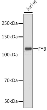
Figure 1. Western blot analysis of FYB using anti-FYB antibody (M32410-1). Electrophoresis was performed on a 5-20% SDS-PAGE gel at 70V (Stacking gel) / 90V (Resolving gel) for 2-3 hours. The sample well of each lane was loaded with 30 ug of sample under reducing conditions. Lane 1: human TF-1 whole cell lysates, Lane 2: human Jurkat whole cell lysates, Lane 3: human HEL whole cell lysates. After electrophoresis, proteins were transferred to a nitrocellulose membrane at 150 mA for 50-90 minutes. Blocked the membrane with 5% non-fat milk/TBS for 1.5 hour at RT. The membrane was incubated with rabbit anti-FYB antigen affinity purified monoclonal antibody (Catalog # M32410-1) at 1:500 overnight at 4°C, then washed with TBS-0.1%Tween 3 times with 5 minutes each and probed with a goat anti-rabbit IgG-HRP secondary antibody at a dilution of 1:500 for 1.5 hour at RT. The signal is developed using an Enhanced Chemiluminescent detection (ECL) kit (Catalog # EK1002) with Tanon 5200 system. A specific band was detected for FYB at approximately 120 kDa. The expected band size for FYB is at 85 kDa.
Figure 1. Western blot analysis of FYB using anti-FYB antibody (M32410-1). Electrophoresis was performed on a 5-20% SDS-PAGE gel at 70V (Stacking gel) / 90V (Resolving gel) for 2-3 hours. The sample well of each lane was loaded with 30 ug of sample under reducing conditions. Lane 1: human TF-1 whole cell lysates, Lane 2: human Jurkat whole cell lysates, Lane 3: human HEL whole cell lysates. After electrophoresis, proteins were transferred to a nitrocellulose membrane at 150 mA for 50-90 minutes. Blocked the membrane with 5% non-fat milk/TBS for 1.5 hour at RT. The membrane was incubated with rabbit anti-FYB antigen affinity purified monoclonal antibody (Catalog # M32410-1) at 1:500 overnight at 4°C, then washed with TBS-0.1%Tween 3 times with 5 minutes each and probed with a goat anti-rabbit IgG-HRP secondary antibody at a dilution of 1:500 for 1.5 hour at RT. The signal is developed using an Enhanced Chemiluminescent detection (ECL) kit (Catalog # EK1002) with Tanon 5200 system. A specific band was detected for FYB at approximately 120 kDa. The expected band size for FYB is at 85 kDa.
Anti-FYB Rabbit Monoclonal Antibody
M32410-1
ApplicationsImmunoFluorescence, Western Blot, ImmunoCytoChemistry, ImmunoHistoChemistry
Product group Antibodies
ReactivityHuman, Mouse
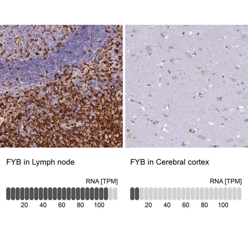
TargetFYB1
Overview
- SupplierBoster Bio
- Product NameAnti-FYB Rabbit Monoclonal Antibody
- Delivery Days Customer9
- ApplicationsImmunoFluorescence, Western Blot, ImmunoCytoChemistry, ImmunoHistoChemistry
- CertificationResearch Use Only
- ClonalityMonoclonal
- Clone ID20F94
- Gene ID2533
- Target nameFYB1
- Target descriptionFYN binding protein 1
- Target synonymsADAP, FYB, PRO0823, SLAP-130, SLAP130, THC3, FYN-binding protein 1, FYB-120/130, FYN-T-binding protein, SLP-76-associated phosphoprotein, adhesion and degranulation-promoting adaptor protein, p120/p130
- HostRabbit
- IsotypeIgG
- Protein IDO15117
- Protein NameFYN-binding protein 1
- Scientific DescriptionBoster Bio Anti-FYB Rabbit Monoclonal Antibody catalog # M32410-1. Tested in WB, IHC, ICC/IF applications. This antibody reacts with Human, Mouse.
- ReactivityHuman, Mouse
- Storage Instruction-20°C
- UNSPSC12352203
Related products
Product group Antibodies
FYB AntibodyCSB-PA002589
ApplicationsWestern Blot, ELISA
ReactivityHuman, Mouse, Rat
TargetFYB1
- SizePrice
Product group Antibodies
Anti-FYB AntibodyA88166
ApplicationsWestern Blot
ReactivityHuman, Mouse
- SizePrice
Product group Antibodies

Goat anti-FYBEB08254
ApplicationsWestern Blot, ELISA
ReactivityBovine, Canine, Human
TargetFYB1
- SizePrice
Product group Antibodies
Anti-FYB AntibodyHPA026796
ApplicationsImmunoHistoChemistry
ReactivityHuman
TargetFYB1
- SizePrice
Product group Antibodies

ApplicationsWestern Blot, ELISA
ReactivityHuman, Mouse, Rat
TargetFYB1
- SizePrice
Product group Antibodies
FYB antibody [N3C3]GTX111564
ApplicationsWestern Blot
ReactivityHuman
TargetFYB1
- SizePrice
Product group Antibodies
ADAP/FYB Recombinant Antibody, AbBy Fluor-555 ConjugatedBSM-61997R-BF555
ApplicationsImmunoFluorescence, Western Blot
ReactivityHuman, Mouse
TargetFYB1
- SizePrice
Product group Antibodies
Anti-FYB (C-term) Antibody102-21081
ApplicationsWestern Blot
TargetFYB1
- SizePrice