


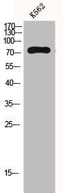
Figure 1. Western blot analysis of GAB2 using anti-GAB2 antibody (A02386-3). Electrophoresis was performed on a 5-20% SDS-PAGE gel at 70V (Stacking gel) / 90V (Resolving gel) for 2-3 hours. The sample well of each lane was loaded with 30 ug of sample under reducing conditions. Lane 1: human K562 whole cell lysates, Lane 2: human SH-SY5Y whole cell lysates, Lane 3: mouse ovary tissue lysates. After electrophoresis, proteins were transferred to a nitrocellulose membrane at 150 mA for 50-90 minutes. Blocked the membrane with 5% non-fat milk/TBS for 1.5 hour at RT. The membrane was incubated with rabbit anti-GAB2 antigen affinity purified polyclonal antibody (Catalog # A02386-3) at 0.25 microg/mL overnight at 4°C, then washed with TBS-0.1%Tween 3 times with 5 minutes each and probed with a goat anti-rabbit IgG-HRP secondary antibody at a dilution of 1:5000 for 1.5 hour at RT. The signal is developed using an Enhanced Chemiluminescent detection (ECL) kit (Catalog # EK1002) with Tanon 5200 system. A specific band was detected for GAB2 at approximately 90 kDa. The expected band size for GAB2 is at 74 kDa.
. Overlay histogram showing HL-60 cells stained with A02386-3 (Blue line). To facilitate intracellular staining, cells were fixed with 4% paraformaldehyde and permeabilized with permeabilization buffer. The cells were blocked with 10% normal goat serum. And then incubated with rabbit anti-GAB2 Antibody (A02386-3, 1 microg/1x106 cells) for 30 min at 20°C. DyLight®488 conjugated goat anti-rabbit IgG (BA1127, 5-10 microg/1x106 cells) was used as secondary antibody for 30 minutes at 20°C. Isotype control antibody (Green line) was rabbit IgG (1 microg/1x106) used under the same conditions. Unlabelled sample without incubation with primary antibody and secondary antibody (Red line) was used as a blank control.")
. Overlay histogram showing HL-60 cells stained with A02386-3 (Blue line). The cells were blocked with 10% normal goat serum. And then incubated with rabbit anti-GAB2 Antibody (A02386-3, 1 microg/1x106 cells) for 30 min at 20°C. PE conjugated goat anti-rabbit IgG (5-10 microg/1x106 cells) was used as secondary antibody for 30 minutes at 20°C. Isotype control antibody (Green line) was rabbit IgG (1 microg/1x106) used under the same conditions. Unlabelled sample (Red line) was also used as a control.")
. Overlay histogram showing THP-1 cells stained with A02386-3 (Blue line). To facilitate intracellular staining, cells were fixed with 4% paraformaldehyde and permeabilized with permeabilization buffer. The cells were blocked with 10% normal goat serum. And then incubated with rabbit anti-GAB2 Antibody (A02386-3, 1 microg/1x106 cells) for 30 min at 20°C. DyLight®488 conjugated goat anti-rabbit IgG (BA1127, 5-10 microg/1x106 cells) was used as secondary antibody for 30 minutes at 20°C. Isotype control antibody (Green line) was rabbit IgG (1 microg/1x106) used under the same conditions. Unlabelled sample without incubation with primary antibody and secondary antibody (Red line) was used as a blank control.")
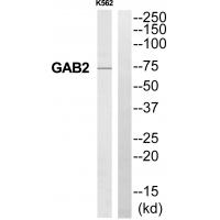
Figure 1. Western blot analysis of GAB2 using anti-GAB2 antibody (A02386-3). Electrophoresis was performed on a 5-20% SDS-PAGE gel at 70V (Stacking gel) / 90V (Resolving gel) for 2-3 hours. The sample well of each lane was loaded with 30 ug of sample under reducing conditions. Lane 1: human K562 whole cell lysates, Lane 2: human SH-SY5Y whole cell lysates, Lane 3: mouse ovary tissue lysates. After electrophoresis, proteins were transferred to a nitrocellulose membrane at 150 mA for 50-90 minutes. Blocked the membrane with 5% non-fat milk/TBS for 1.5 hour at RT. The membrane was incubated with rabbit anti-GAB2 antigen affinity purified polyclonal antibody (Catalog # A02386-3) at 0.25 microg/mL overnight at 4°C, then washed with TBS-0.1%Tween 3 times with 5 minutes each and probed with a goat anti-rabbit IgG-HRP secondary antibody at a dilution of 1:5000 for 1.5 hour at RT. The signal is developed using an Enhanced Chemiluminescent detection (ECL) kit (Catalog # EK1002) with Tanon 5200 system. A specific band was detected for GAB2 at approximately 90 kDa. The expected band size for GAB2 is at 74 kDa.
Anti-GAB2 Antibody Picoband(r)
A02386-3-CARRIER-FREE
ApplicationsFlow Cytometry, Western Blot, ELISA
Product group Antibodies
ReactivityHuman, Mouse
TargetGAB2
Overview
- SupplierBoster Bio
- Product NameAnti-GAB2 Antibody Picoband(r)
- Delivery Days Customer9
- ApplicationsFlow Cytometry, Western Blot, ELISA
- CertificationResearch Use Only
- ClonalityPolyclonal
- Concentration500 ug/ml
- Gene ID9846
- Target nameGAB2
- Target descriptionGRB2 associated binding protein 2
- Target synonymsGRB2-associated-binding protein 2, Grb2-associated binder 2, growth factor receptor bound protein 2-associated protein 2, pp100
- HostRabbit
- IsotypeIgG
- Protein IDQ9UQC2
- Protein NameGRB2-associated-binding protein 2
- Scientific DescriptionBoster Bio Anti-GAB2 Antibody Picoband® catalog # A02386-3. Tested in ELISA, Flow Cytometry, WB applications. This antibody reacts with Human, Mouse. The brand Picoband indicates this is a premium antibody that guarantees superior quality, high affinity, and strong signals with minimal background in Western blot applications. Only our best-performing antibodies are designated as Picoband, ensuring unmatched performance.
- ReactivityHuman, Mouse
- Storage Instruction-20°C,2°C to 8°C
- UNSPSC12352203
Related products
Product group Antibodies
GAB2 AntibodyCSB-PA002610
ApplicationsWestern Blot, ELISA
ReactivityHuman, Mouse, Rat
TargetGAB2
- SizePrice
Product group Antibodies

GRB2 (Phospho-Ser159) AntibodyABX012651
ApplicationsWestern Blot, ELISA, ImmunoHistoChemistry
- SizePrice
Product group Antibodies
Anti-GAB2 AntibodyA43782
ApplicationsWestern Blot
ReactivityHuman, Mouse, Rat
- SizePrice
Product group Antibodies

Goat anti-GAB2EB07759
ApplicationsWestern Blot, ELISA
ReactivityCanine, Human, Mouse, Rat
TargetGAB2
- SizePrice

Product group Antibodies
Anti-GAB2 AntibodyHPA000271
ApplicationsImmunoHistoChemistry
ReactivityHuman
TargetGAB2
- SizePrice
Product group Antibodies

GAB2 AntibodyLS-C401696
ApplicationsWestern Blot, ELISA, ImmunoHistoChemistry
ReactivityHuman, Mouse, Rat
TargetGAB2
- SizePrice
Product group Antibodies
Gab2 Polyclonal AntibodyCAC09951
ApplicationsImmunoFluorescence, Western Blot, ELISA, ImmunoHistoChemistry
ReactivityMouse
TargetGAB2
- SizePrice
Product group Antibodies
GAB4/GAB2 AntibodyPACO03441
ApplicationsWestern Blot, ELISA, ImmunoHistoChemistry
ReactivityHuman
TargetGAB2
- SizePrice
Product group Antibodies
References
Gab2 Polyclonal AntibodyBS-2885R
ApplicationsFlow Cytometry, ImmunoFluorescence, Western Blot, ELISA, ImmunoCytoChemistry, ImmunoHistoChemistry, ImmunoHistoChemistry Frozen, ImmunoHistoChemistry Paraffin
ReactivityBovine, Canine, Equine, Human, Mouse, Porcine, Rabbit, Rat
TargetGAB2
- SizePrice
![Various tissue extracts (50 μg) were separated by 7.5% SDS-PAGE, and the membrane was blotted with GAB2 antibody [N1C1] (GTX114226) diluted at 1:1000. The HRP-conjugated anti-rabbit IgG antibody (GTX213110-01) was used to detect the primary antibody.](https://www.genetex.com/upload/website/prouct_img/normal/GTX114226/GTX114226_40625_20210416_WB_M_R_w_23060501_934.webp)
Product group Antibodies
GAB2 antibody [N1C1]GTX114226
ApplicationsWestern Blot
ReactivityHuman, Mouse, Rat
TargetGAB2
- SizePrice