


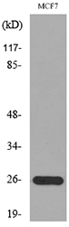
Figure 1. Western blot analysis of Galectin 3 using anti-Galectin 3 antibody (M00621-4). Electrophoresis was performed on a 5-20% SDS-PAGE gel at 70V (Stacking gel) / 90V (Resolving gel) for 2-3 hours. The sample well of each lane was loaded with 30 ug of sample under reducing conditions. Lane 1: human A431 whole cell lysates, Lane 2: human SIHA whole cell lysates, Lane 3: human RT4 whole cell lysates, Lane 4: human MCF-7 whole cell lysates, Lane 5: rat PC-12 whole cell lysates. After electrophoresis, proteins were transferred to a nitrocellulose membrane at 150 mA for 50-90 minutes. Blocked the membrane with 5% non-fat milk/TBS for 1.5 hour at RT. The membrane was incubated with rabbit anti-Galectin 3 antigen affinity purified monoclonal antibody (Catalog # M00621-4) at 1:500 overnight at 4°C, then washed with TBS-0.1%Tween 3 times with 5 minutes each and probed with a goat anti-rabbit IgG-HRP secondary antibody at a dilution of 1:5000 for 1.5 hour at RT. The signal is developed using an Enhanced Chemiluminescent detection (ECL) kit (Catalog # EK1002) with Tanon 5200 system. A specific band was detected for Galectin 3 at approximately 29 kDa. The expected band size for Galectin 3 is at 26 kDa.
Figure 1. Western blot analysis of Galectin 3 using anti-Galectin 3 antibody (M00621-4). Electrophoresis was performed on a 5-20% SDS-PAGE gel at 70V (Stacking gel) / 90V (Resolving gel) for 2-3 hours. The sample well of each lane was loaded with 30 ug of sample under reducing conditions. Lane 1: human A431 whole cell lysates, Lane 2: human SIHA whole cell lysates, Lane 3: human RT4 whole cell lysates, Lane 4: human MCF-7 whole cell lysates, Lane 5: rat PC-12 whole cell lysates. After electrophoresis, proteins were transferred to a nitrocellulose membrane at 150 mA for 50-90 minutes. Blocked the membrane with 5% non-fat milk/TBS for 1.5 hour at RT. The membrane was incubated with rabbit anti-Galectin 3 antigen affinity purified monoclonal antibody (Catalog # M00621-4) at 1:500 overnight at 4°C, then washed with TBS-0.1%Tween 3 times with 5 minutes each and probed with a goat anti-rabbit IgG-HRP secondary antibody at a dilution of 1:5000 for 1.5 hour at RT. The signal is developed using an Enhanced Chemiluminescent detection (ECL) kit (Catalog # EK1002) with Tanon 5200 system. A specific band was detected for Galectin 3 at approximately 29 kDa. The expected band size for Galectin 3 is at 26 kDa.
Anti-Galectin 3 Rabbit Monoclonal Antibody
M00621-4
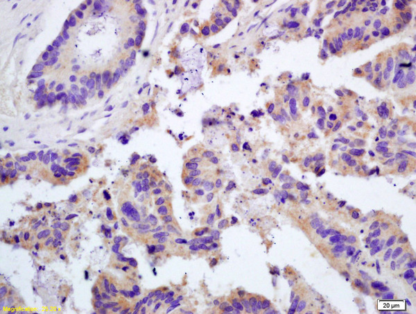
ApplicationsFlow Cytometry, ImmunoFluorescence, Western Blot, ImmunoCytoChemistry, ImmunoHistoChemistry
Product group Antibodies
ReactivityHuman, Mouse
TargetLGALS3
Overview
- SupplierBoster Bio
- Product NameAnti-Galectin 3 Rabbit Monoclonal Antibody
- Delivery Days Customer9
- ApplicationsFlow Cytometry, ImmunoFluorescence, Western Blot, ImmunoCytoChemistry, ImmunoHistoChemistry
- CertificationResearch Use Only
- ClonalityMonoclonal
- Clone ID28L32
- Gene ID3958
- Target nameLGALS3
- Target descriptiongalectin 3
- Target synonymsCBP35, GAL3, GALBP, GALIG, L31, LGALS2, MAC2, galectin-3, 35 kDa lectin, IgE-binding protein, MAC-2 antigen, advanced glycation end-product receptor 3, carbohydrate-binding protein 35, epididymis secretory sperm binding protein, galactose-specific lectin 3, laminin-binding protein, lectin L-29, lectin, galactoside-binding, soluble, 3
- HostRabbit
- IsotypeIgG
- Protein IDP17931
- Protein NameGalectin-3
- Scientific DescriptionBoster Bio Anti-Galectin 3 Rabbit Monoclonal Antibody catalog # M00621-4. Tested in WB, IHC, ICC/IF, Flow Cytometry applications. This antibody reacts with Human, Mouse.
- ReactivityHuman, Mouse
- Storage Instruction-20°C
- UNSPSC12352203
References
- Shi J, Wang Z, Wu B, et al. Cofilin-1, peroxiredoxin-1, and galectin-3: Major proteins released by macrophages infected with Corynebacterium pseudotuberculosis. Vet Microbiol. 2019,239:108461. doi: 10.1016/j.vetmic.2019.108461Read this paper
Related products
Product group Antibodies
ReactivityHuman
TargetLGALS3
- SizePrice
Product group Antibodies
LGALS3 Polyclonal AntibodyCAC13799
ApplicationsWestern Blot, ELISA, ImmunoHistoChemistry
TargetLGALS3
- SizePrice
Product group Antibodies
Anti-Galectin-3 Antibody130-10090
ApplicationsELISA
ReactivityHuman
TargetLGALS3
- SizePrice
Product group Antibodies
References
Galectin 3 Polyclonal AntibodyBS-0721R
ApplicationsFlow Cytometry, ImmunoFluorescence, ELISA, ImmunoCytoChemistry, ImmunoHistoChemistry, ImmunoHistoChemistry Frozen, ImmunoHistoChemistry Paraffin
ReactivityHuman, Mouse
TargetLGALS3
- SizePrice

Product group Antibodies
ApplicationsImmunoCytoChemistry
ReactivityHuman
TargetLGALS3
- SizePrice
Product group Antibodies
Anti-LGALS3 AntibodyA100515
ApplicationsWestern Blot, ELISA
ReactivityHuman
- SizePrice
Product group Antibodies

ApplicationsWestern Blot, ELISA
ReactivityHuman, Mouse, Rat
TargetLGALS3
- SizePrice
Product group Antibodies
References
Galectin 3 antibodyGTX113486
ApplicationsImmunoFluorescence, Western Blot, ELISA, ImmunoCytoChemistry, ImmunoHistoChemistry, ImmunoHistoChemistry Paraffin
ReactivityCanine, Human, Rat
TargetLGALS3
- SizePrice