


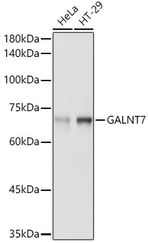
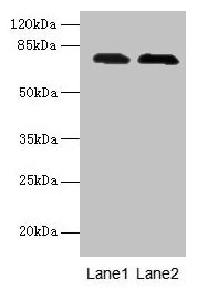
Figure 1. Western blot analysis of GALNT7 using anti-GALNT7 antibody (A10021-1). Electrophoresis was performed on a 5-20% SDS-PAGE gel at 70V (Stacking gel) / 90V (Resolving gel) for 2-3 hours. The sample well of each lane was loaded with 30 ug of sample under reducing conditions. Lane 1: human Hela whole cell lysates, Lane 2: human HepG2 whole cell lysates, Lane 3: human A431 whole cell lysates, Lane 4: human Hacat whole cell lysates, Lane 5: rat brain tissue lysates, Lane 6: rat kidney tissue lysates, Lane 7: mouse brain tissue lysates, Lane 8: mouse kidney tissue lysates. After electrophoresis, proteins were transferred to a nitrocellulose membrane at 150 mA for 50-90 minutes. Blocked the membrane with 5% non-fat milk/TBS for 1.5 hour at RT. The membrane was incubated with rabbit anti-GALNT7 antigen affinity purified polyclonal antibody (Catalog # A10021-1) at 0.5 microg/mL overnight at 4°C, then washed with TBS-0.1%Tween 3 times with 5 minutes each and probed with a goat anti-rabbit IgG-HRP secondary antibody at a dilution of 1:5000 for 1.5 hour at RT. The signal is developed using an Enhanced Chemiluminescent detection (ECL) kit (Catalog # EK1002) with Tanon 5200 system. A specific band was detected for GALNT7 at approximately 75 kDa. The expected band size for GALNT7 is at 75 kDa.
Figure 1. Western blot analysis of GALNT7 using anti-GALNT7 antibody (A10021-1). Electrophoresis was performed on a 5-20% SDS-PAGE gel at 70V (Stacking gel) / 90V (Resolving gel) for 2-3 hours. The sample well of each lane was loaded with 30 ug of sample under reducing conditions. Lane 1: human Hela whole cell lysates, Lane 2: human HepG2 whole cell lysates, Lane 3: human A431 whole cell lysates, Lane 4: human Hacat whole cell lysates, Lane 5: rat brain tissue lysates, Lane 6: rat kidney tissue lysates, Lane 7: mouse brain tissue lysates, Lane 8: mouse kidney tissue lysates. After electrophoresis, proteins were transferred to a nitrocellulose membrane at 150 mA for 50-90 minutes. Blocked the membrane with 5% non-fat milk/TBS for 1.5 hour at RT. The membrane was incubated with rabbit anti-GALNT7 antigen affinity purified polyclonal antibody (Catalog # A10021-1) at 0.5 microg/mL overnight at 4°C, then washed with TBS-0.1%Tween 3 times with 5 minutes each and probed with a goat anti-rabbit IgG-HRP secondary antibody at a dilution of 1:5000 for 1.5 hour at RT. The signal is developed using an Enhanced Chemiluminescent detection (ECL) kit (Catalog # EK1002) with Tanon 5200 system. A specific band was detected for GALNT7 at approximately 75 kDa. The expected band size for GALNT7 is at 75 kDa.
Anti-GALNT7 Antibody Picoband(r)
A10021-1-DYLIGHT488
ApplicationsWestern Blot, ELISA
Product group Antibodies
ReactivityHuman, Mouse, Rat
TargetGALNT7
Overview
- SupplierBoster Bio
- Product NameAnti-GALNT7 Antibody Picoband(r)
- Delivery Days Customer9
- ApplicationsWestern Blot, ELISA
- CertificationResearch Use Only
- ClonalityPolyclonal
- Concentration500 ug/ml
- ConjugateDyLight 488
- Gene ID51809
- Target nameGALNT7
- Target descriptionpolypeptide N-acetylgalactosaminyltransferase 7
- Target synonymsGALNAC-T7, GalNAcT7, N-acetylgalactosaminyltransferase 7, UDP-GalNAc:polypeptide N-acetylgalactosaminyltransferase 7, UDP-N-acetyl-alpha-D-galactosamine:polypeptide N-acetylgalactosaminyltransferase 7 (GalNAc-T7), polypeptide GalNAc transferase 7, pp-GaNTase 7, protein-UDP acetylgalactosaminyltransferase 7
- HostRabbit
- Protein IDQ86SF2
- Protein NameN-acetylgalactosaminyltransferase 7
- Scientific DescriptionBoster Bio Anti-GALNT7 Antibody Picoband® catalog # A10021-1. Tested in WB, ELISA applications. This antibody reacts with Human, Mouse, Rat. The brand Picoband indicates this is a premium antibody that guarantees superior quality, high affinity, and strong signals with minimal background in Western blot applications. Only our best-performing antibodies are designated as Picoband, ensuring unmatched performance.
- ReactivityHuman, Mouse, Rat
- Storage Instruction-20°C,2°C to 8°C
- UNSPSC12352203
Related products
Product group Antibodies
Galnt7 Polyclonal AntibodyCAC09438
ApplicationsWestern Blot, ELISA, ImmunoHistoChemistry
TargetGALNT7
- SizePrice
Product group Antibodies
Anti-GALNT7 AntibodyA308233
ApplicationsWestern Blot, ImmunoHistoChemistry
ReactivityHuman, Mouse, Rat
- SizePrice
Product group Antibodies
Anti-GALNT7 Antibody107-10582
ApplicationsImmunoFluorescence, Western Blot, ImmunoCytoChemistry, ImmunoHistoChemistry, ImmunoHistoChemistry Paraffin
ReactivityHuman, Mouse
TargetGALNT7
- SizePrice
Product group Antibodies
References
GALNT7 antibody [N1C2]GTX106068
ApplicationsImmunoFluorescence, Western Blot, ImmunoCytoChemistry, ImmunoHistoChemistry, ImmunoHistoChemistry Paraffin
ReactivityHuman, Mouse, Rat
TargetGALNT7
- SizePrice
Product group Antibodies

GALNT7 AntibodyLS-C398978
ApplicationsWestern Blot, ELISA, ImmunoHistoChemistry, ImmunoHistoChemistry Paraffin
ReactivityHuman
TargetGALNT7
- SizePrice

Product group Antibodies
Anti-GALNT7 AntibodyHPA065317
ApplicationsImmunoCytoChemistry, ImmunoHistoChemistry
ReactivityHuman
TargetGALNT7
- SizePrice
Product group Antibodies
GALNT7 AntibodyCSB-PA773016LA01HU
ApplicationsWestern Blot, ELISA, ImmunoHistoChemistry
ReactivityHuman
TargetGALNT7
- SizePrice
Product group Antibodies
Anti-GALNT7Y058485
ApplicationsWestern Blot, ImmunoHistoChemistry
ReactivityHuman
- SizePrice
Product group Antibodies
Anti-GALNT7 Antibody Picoband(r)A10021-1-CARRIER-FREE
ApplicationsWestern Blot, ELISA
ReactivityHuman, Mouse, Rat
TargetGALNT7
- SizePrice