


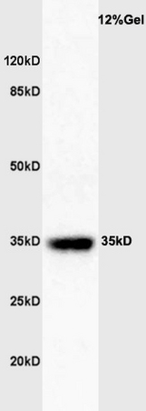
Figure 1. Western blot analysis of GATA1 using anti-GATA1 antibody (A00408-2). Electrophoresis was performed on a 5-20% SDS-PAGE gel at 70V (Stacking gel) / 90V (Resolving gel) for 2-3 hours. The sample well of each lane was loaded with 50ug of sample under reducing conditions. Lane 1: human K562 whole cell lysates. After Electrophoresis, proteins were transferred to a Nitrocellulose membrane at 150mA for 50-90 minutes. Blocked the membrane with 5% Non-fat Milk/ TBS for 1.5 hour at RT. The membrane was incubated with rabbit anti-GATA1 antigen affinity purified polyclonal antibody (Catalog # A00408-2) at 0.5 microg/mL overnight at 4°C, then washed with TBS-0.1%Tween 3 times with 5 minutes each and probed with a goat anti-rabbit IgG-HRP secondary antibody at a dilution of 1:5000 for 1.5 hour at RT. The signal is developed using an Enhanced Chemiluminescent detection (ECL) kit (Catalog # EK1002) with Tanon 5200 system. A specific band was detected for GATA1 at approximately 45KD. The expected band size for GATA1 is at 45KD.
. Overlay histogram showing K562 cells stained with A00408-2 (Blue line). To facilitate intracellular staining, cells were fixed with 4% paraformaldehyde and permeabilized with permeabilization buffer. The cells were blocked with 10% normal goat serum. And then incubated with rabbit anti- GATA1 Antibody (A00408-2, 1microg/1x106 cells) for 30 min at 20°C. DyLight®488 conjugated goat anti-rabbit IgG (BA1127, 5-10microg/1x106 cells) was used as secondary antibody for 30 minutes at 20°C. Isotype control antibody (Green line) was rabbit IgG (1microg/1x106) used under the same conditions. Unlabelled sample without incubation with primary antibody and secondary antibody (Red line) was used as a blank control.")
Figure 1. Western blot analysis of GATA1 using anti-GATA1 antibody (A00408-2). Electrophoresis was performed on a 5-20% SDS-PAGE gel at 70V (Stacking gel) / 90V (Resolving gel) for 2-3 hours. The sample well of each lane was loaded with 50ug of sample under reducing conditions. Lane 1: human K562 whole cell lysates. After Electrophoresis, proteins were transferred to a Nitrocellulose membrane at 150mA for 50-90 minutes. Blocked the membrane with 5% Non-fat Milk/ TBS for 1.5 hour at RT. The membrane was incubated with rabbit anti-GATA1 antigen affinity purified polyclonal antibody (Catalog # A00408-2) at 0.5 microg/mL overnight at 4°C, then washed with TBS-0.1%Tween 3 times with 5 minutes each and probed with a goat anti-rabbit IgG-HRP secondary antibody at a dilution of 1:5000 for 1.5 hour at RT. The signal is developed using an Enhanced Chemiluminescent detection (ECL) kit (Catalog # EK1002) with Tanon 5200 system. A specific band was detected for GATA1 at approximately 45KD. The expected band size for GATA1 is at 45KD.
Anti-GATA1 Antibody Picoband(r)
A00408-2-PE
ApplicationsFlow Cytometry, Western Blot, ELISA
Product group Antibodies
ReactivityHuman
TargetGATA1
Overview
- SupplierBoster Bio
- Product NameAnti-GATA1 Antibody Picoband(r)
- Delivery Days Customer9
- ApplicationsFlow Cytometry, Western Blot, ELISA
- CertificationResearch Use Only
- ClonalityPolyclonal
- Concentration500 ug/ml
- ConjugateRPE
- Gene ID2623
- Target nameGATA1
- Target descriptionGATA binding protein 1
- Target synonymsCNSHA9, ERYF1, GATA-1, GF-1, GF1, HAEADA, NF-E1, NFE1, XLANP, XLTDA, XLTT, erythroid transcription factor, GATA-binding factor 1, NF-E1 DNA-binding protein, erythroid transcription factor 1, globin transcription factor 1, nuclear factor, erythroid 1, transcription factor GATA1
- HostRabbit
- IsotypeIgG
- Protein IDP15976
- Protein NameErythroid transcription factor
- Scientific DescriptionBoster Bio Anti-GATA1 Antibody Picoband® catalog # A00408-2. Tested in ELISA, Flow Cytometry, WB applications. This antibody reacts with Human. The brand Picoband indicates this is a premium antibody that guarantees superior quality, high affinity, and strong signals with minimal background in Western blot applications. Only our best-performing antibodies are designated as Picoband, ensuring unmatched performance.
- ReactivityHuman
- Storage Instruction-20°C,2°C to 8°C
- UNSPSC12352203
Related products
Product group Antibodies
GATA1 Polyclonal AntibodyBS-3872R
ApplicationsFlow Cytometry, ImmunoFluorescence, Western Blot, ELISA, ImmunoCytoChemistry, ImmunoHistoChemistry, ImmunoHistoChemistry Frozen, ImmunoHistoChemistry Paraffin
ReactivityBovine, Canine, Equine, Human, Mouse, Porcine, Rabbit, Rat
TargetGATA1
- SizePrice
Product group Antibodies
GATA1 Polyclonal AntibodyCAC14535
ApplicationsWestern Blot, ELISA, ImmunoHistoChemistry
TargetGATA1
- SizePrice
Product group Antibodies
Anti-GATA1 Antibody101-10607
ApplicationsWestern Blot, ELISA
TargetGATA1
- SizePrice
Product group Antibodies
Anti-GATA1 AntibodyA121166
ApplicationsFlow Cytometry, ImmunoFluorescence, Western Blot, ELISA
ReactivityHuman
- SizePrice
Product group Antibodies

Goat anti-GATA1 AntibodyEB05571
ApplicationsFlow Cytometry, ImmunoFluorescence, Western Blot, ELISA
ReactivityHuman
TargetGATA1
- SizePrice
Product group Antibodies
References
GATA1 antibody [N1C1]GTX113193
ApplicationsWestern Blot
ReactivityHuman
TargetGATA1
- SizePrice
Product group Antibodies

GATA1 Antibody (phospho-Ser142)LS-C358759
ApplicationsImmunoPrecipitation, Western Blot, ImmunoHistoChemistry, ImmunoHistoChemistry Paraffin
ReactivityCanine, Human, Monkey, Mouse, Porcine, Rat
TargetGATA1
- SizePrice