

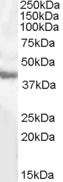
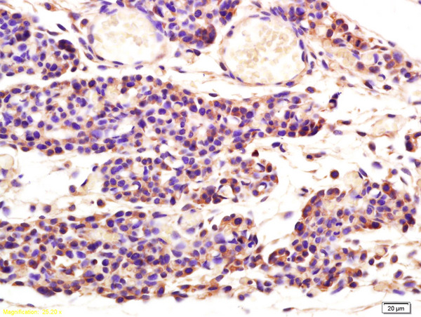
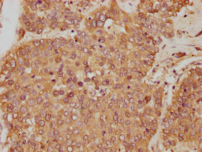
Anti-GOT1 Antibody
ER1802-48
ApplicationsFlow Cytometry, Western Blot, ImmunoHistoChemistry, ImmunoHistoChemistry Paraffin
Product group Antibodies
ReactivityHuman
TargetGOT1
Overview
- SupplierHUABIO
- Product NameAnti-GOT1 Antibody
- Delivery Days Customer7
- ApplicationsFlow Cytometry, Western Blot, ImmunoHistoChemistry, ImmunoHistoChemistry Paraffin
- Applications SupplierWB,IHC-P,FC
- CertificationResearch Use Only
- ClonalityPolyclonal
- Concentration1 mg/ml
- ConjugateUnconjugated
- Gene ID2805
- Target nameGOT1
- Target descriptionglutamic-oxaloacetic transaminase 1
- Target synonymsAST, AST1, ASTQTL1, GIG18, SGOT, cAspAT, cCAT, aspartate aminotransferase, cytoplasmic, aspartate aminotransferase 1, aspartate transaminase 1, cysteine aminotransferase, cytoplasmic, cysteine transaminase, cytoplasmic, glutamate oxaloacetate transaminase 1, glutamic-oxaloacetic transaminase 1, soluble, growth-inhibiting protein 18, testis secretory sperm-binding protein Li 196a, transaminase A
- HostRabbit
- IsotypeIgG
- Protein IDP17174
- Protein NameAspartate aminotransferase, cytoplasmic
- Scientific DescriptionBiosynthesis of L-glutamate from L-aspartate or L-cysteine. Important regulator of levels of glutamate, the major excitatory neurotransmitter of the vertebrate central nervous system. Acts as a scavenger of glutamate in brain neuroprotection. The aspartate aminotransferase activity is involved in hepatic glucose synthesis during development and in adipocyte glyceroneogenesis. Using L-cysteine as substrate, regulates levels of mercaptopyruvate, an important source of hydrogen sulfide. Mercaptopyruvate is converted into H2S via the action of 3-mercaptopyruvate sulfurtransferase (3MST). Hydrogen sulfide is an important synaptic modulator and neuroprotectant in the brain.
- ReactivityHuman
- Reactivity SupplierHuman
- Storage Instruction-20°C,2°C to 8°C
- UNSPSC41116161
Datasheet
Related products
Product group Antibodies
ApplicationsWestern Blot, ELISA
ReactivityHuman
- SizePrice

Product group Antibodies
Anti-Aspartate Aminotransferase/GOT1 Antibody Picoband(r)A04085-3-CARRIER-FREE
ApplicationsFlow Cytometry, ImmunoFluorescence, Western Blot, ELISA, ImmunoCytoChemistry, ImmunoHistoChemistry
ReactivityHuman, Mouse, Rat
TargetGOT1
- SizePrice
Product group Antibodies
Anti-GOT1 Antibody144-05822
ApplicationsImmunoFluorescence, Western Blot, ImmunoHistoChemistry
ReactivityHuman, Mouse, Rat
TargetGOT1
- SizePrice
Product group Antibodies
ApplicationsImmunoFluorescence, Western Blot, ELISA, ImmunoCytoChemistry, ImmunoHistoChemistry, ImmunoHistoChemistry Frozen, ImmunoHistoChemistry Paraffin
ReactivityBovine, Canine, Human, Mouse, Porcine, Rabbit, Rat
TargetGOT1
- SizePrice
Product group Antibodies
GOT1 AntibodyCSB-PA009679HA01HU
ApplicationsImmunoFluorescence, Western Blot, ELISA, ImmunoHistoChemistry
ReactivityHuman, Mouse
TargetGOT1
- SizePrice
Product group Antibodies

Goat anti-GOT1 (aa 22-35)EB08132
ApplicationsWestern Blot, ELISA
ReactivityHuman, Mouse
TargetGOT1
- SizePrice
Product group Antibodies
GOT1 Polyclonal AntibodyCAC13746
ApplicationsImmunoFluorescence, Western Blot, ELISA, ImmunoHistoChemistry
ReactivityMouse
TargetGOT1
- SizePrice
Product group Antibodies

Aspartate Aminotransferase AntibodyLS-C404720
ApplicationsWestern Blot, ELISA, ImmunoHistoChemistry
ReactivityHuman, Mouse, Rat
TargetGOT1
- SizePrice

Product group Antibodies
Anti-GOT1 AntibodyHPA064532
ApplicationsWestern Blot, ImmunoHistoChemistry
ReactivityHuman
TargetGOT1
- SizePrice
![Various tissue extracts (30 μg) were separated by 10% SDS-PAGE, and the membrane was blotted with GOT1 antibody [GT638] (GTX632031) diluted at 1:5000. The HRP-conjugated anti-mouse IgG antibody (GTX213111-01) was used to detect the primary antibody.](https://www.genetex.com/upload/website/prouct_img/normal/GTX632031/GTX632031_41918_20180622_WB_M_tissue_w_23061202_960.webp)
Product group Antibodies
GOT1 antibody [GT638]GTX632031
ApplicationsWestern Blot
ReactivityHuman, Mouse
TargetGOT1
- SizePrice