


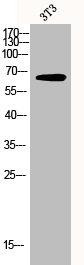
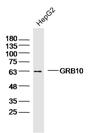
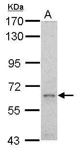
Figure 1. Western blot analysis of GRB10 using anti-GRB10 antibody (A01663-2). Electrophoresis was performed on a 5-20% SDS-PAGE gel at 70V (Stacking gel) / 90V (Resolving gel) for 2-3 hours. The sample well of each lane was loaded with 30 ug of sample under reducing conditions. Lane 1: human Hela whole cell lysates, Lane 2: human HepG2 whole cell lysates, Lane 3: human 293T whole cell lysates, Lane 4: monkey COS-7 whole cell lysates, Lane 5: human K562 whole cell lysates, Lane 6: human U87 whole cell lysates, Lane 7: human HEL whole cell lysates, Lane 8: rat liver tissue lysates, Lane 9: rat brain tissue lysates, Lane 10: mouse liver tissue lysates, Lane 11: mouse brain tissue lysates. After electrophoresis, proteins were transferred to a nitrocellulose membrane at 150 mA for 50-90 minutes. Blocked the membrane with 5% non-fat milk/TBS for 1.5 hour at RT. The membrane was incubated with rabbit anti-GRB10 antigen affinity purified polyclonal antibody (Catalog # A01663-2) at 0.5 microg/mL overnight at 4°C, then washed with TBS-0.1%Tween 3 times with 5 minutes each and probed with a goat anti-rabbit IgG-HRP secondary antibody at a dilution of 1:5000 for 1.5 hour at RT. The signal is developed using an Enhanced Chemiluminescent detection (ECL) kit (Catalog # EK1002) with Tanon 5200 system. A specific band was detected for GRB10 at approximately 76 kDa. The expected band size for GRB10 is at 67 kDa.
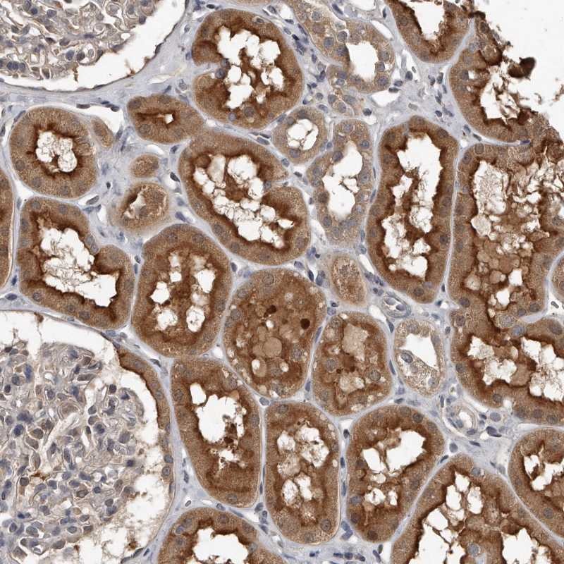
. GRB10 was detected in a paraffin-embedded section of human colorectal adenocarcinoma tissue. Heat mediated antigen retrieval was performed in EDTA buffer (pH 8.0, epitope retrieval solution). The tissue section was blocked with 10% goat serum. The tissue section was then incubated with 2 microg/ml rabbit anti-GRB10 Antibody (A01663-2) overnight at 4°C. Peroxidase Conjugated Goat Anti-rabbit IgG was used as secondary antibody and incubated for 30 minutes at 37°C. The tissue section was developed using HRP Conjugated Rabbit IgG Super Vision Assay Kit (Catalog # SV0002) with DAB as the chromogen.")
. GRB10 was detected in a paraffin-embedded section of human laryngeal squamous cell carcinoma tissue. Heat mediated antigen retrieval was performed in EDTA buffer (pH 8.0, epitope retrieval solution). The tissue section was blocked with 10% goat serum. The tissue section was then incubated with 2 microg/ml rabbit anti-GRB10 Antibody (A01663-2) overnight at 4°C. Peroxidase Conjugated Goat Anti-rabbit IgG was used as secondary antibody and incubated for 30 minutes at 37°C. The tissue section was developed using HRP Conjugated Rabbit IgG Super Vision Assay Kit (Catalog # SV0002) with DAB as the chromogen.")
. GRB10 was detected in a paraffin-embedded section of human liver cancer tissue. Heat mediated antigen retrieval was performed in EDTA buffer (pH 8.0, epitope retrieval solution). The tissue section was blocked with 10% goat serum. The tissue section was then incubated with 2 microg/ml rabbit anti-GRB10 Antibody (A01663-2) overnight at 4°C. Peroxidase Conjugated Goat Anti-rabbit IgG was used as secondary antibody and incubated for 30 minutes at 37°C. The tissue section was developed using HRP Conjugated Rabbit IgG Super Vision Assay Kit (Catalog # SV0002) with DAB as the chromogen.")
. GRB10 was detected in a paraffin-embedded section of mouse brain tissue. Heat mediated antigen retrieval was performed in EDTA buffer (pH 8.0, epitope retrieval solution). The tissue section was blocked with 10% goat serum. The tissue section was then incubated with 2 microg/ml rabbit anti-GRB10 Antibody (A01663-2) overnight at 4°C. Peroxidase Conjugated Goat Anti-rabbit IgG was used as secondary antibody and incubated for 30 minutes at 37°C. The tissue section was developed using HRP Conjugated Rabbit IgG Super Vision Assay Kit (Catalog # SV0002) with DAB as the chromogen.")
. Overlay histogram showing U87 cells stained with A01663-2 (Blue line). To facilitate intracellular staining, cells were fixed with 4% paraformaldehyde and permeabilized with permeabilization buffer. The cells were blocked with 10% normal goat serum. And then incubated with rabbit anti-GRB10 Antibody (A01663-2, 1 microg/1x106 cells) for 30 min at 20°C. DyLight®488 conjugated goat anti-rabbit IgG (BA1127, 5-10 microg/1x106 cells) was used as secondary antibody for 30 minutes at 20°C. Isotype control antibody (Green line) was rabbit IgG (1 microg/1x106) used under the same conditions. Unlabelled sample (Red line) was also used as a control.")
Figure 1. Western blot analysis of GRB10 using anti-GRB10 antibody (A01663-2). Electrophoresis was performed on a 5-20% SDS-PAGE gel at 70V (Stacking gel) / 90V (Resolving gel) for 2-3 hours. The sample well of each lane was loaded with 30 ug of sample under reducing conditions. Lane 1: human Hela whole cell lysates, Lane 2: human HepG2 whole cell lysates, Lane 3: human 293T whole cell lysates, Lane 4: monkey COS-7 whole cell lysates, Lane 5: human K562 whole cell lysates, Lane 6: human U87 whole cell lysates, Lane 7: human HEL whole cell lysates, Lane 8: rat liver tissue lysates, Lane 9: rat brain tissue lysates, Lane 10: mouse liver tissue lysates, Lane 11: mouse brain tissue lysates. After electrophoresis, proteins were transferred to a nitrocellulose membrane at 150 mA for 50-90 minutes. Blocked the membrane with 5% non-fat milk/TBS for 1.5 hour at RT. The membrane was incubated with rabbit anti-GRB10 antigen affinity purified polyclonal antibody (Catalog # A01663-2) at 0.5 microg/mL overnight at 4°C, then washed with TBS-0.1%Tween 3 times with 5 minutes each and probed with a goat anti-rabbit IgG-HRP secondary antibody at a dilution of 1:5000 for 1.5 hour at RT. The signal is developed using an Enhanced Chemiluminescent detection (ECL) kit (Catalog # EK1002) with Tanon 5200 system. A specific band was detected for GRB10 at approximately 76 kDa. The expected band size for GRB10 is at 67 kDa.
Anti-GRB10 Antibody Picoband(r)
A01663-2-CARRIER-FREE
ApplicationsFlow Cytometry, Western Blot, ELISA, ImmunoHistoChemistry
Product group Antibodies
ReactivityHuman, Monkey, Mouse, Rat
TargetGRB10
Overview
- SupplierBoster Bio
- Product NameAnti-GRB10 Antibody Picoband(r)
- Delivery Days Customer9
- ApplicationsFlow Cytometry, Western Blot, ELISA, ImmunoHistoChemistry
- CertificationResearch Use Only
- ClonalityPolyclonal
- Concentration500 ug/ml
- Gene ID2887
- Target nameGRB10
- Target descriptiongrowth factor receptor bound protein 10
- Target synonymsGRB-IR, Grb-10, IRBP, MEG1, RSS, growth factor receptor-bound protein 10, GRB10 adapter protein, GRB10 adaptor protein, insulin receptor-binding protein Grb-IR, maternally expressed gene 1
- HostRabbit
- IsotypeIgG
- Protein IDQ13322
- Protein NameGrowth factor receptor-bound protein 10
- Scientific DescriptionBoster Bio Anti-GRB10 Antibody Picoband® catalog # A01663-2. Tested in ELISA, Flow Cytometry, IHC, WB applications. This antibody reacts with Human, Monkey, Mouse, Rat. The brand Picoband indicates this is a premium antibody that guarantees superior quality, high affinity, and strong signals with minimal background in Western blot applications. Only our best-performing antibodies are designated as Picoband, ensuring unmatched performance.
- ReactivityHuman, Monkey, Mouse, Rat
- Storage Instruction-20°C,2°C to 8°C
- UNSPSC12352203
Related products
Product group Antibodies
GRB10 AntibodyCSB-PA002806
ApplicationsImmunoFluorescence, Western Blot, ELISA, ImmunoHistoChemistry
ReactivityHuman
TargetGRB10
- SizePrice

Product group Antibodies
Anti-GRB10 AntibodyA83162
ApplicationsWestern Blot, ELISA
ReactivityHuman
- SizePrice
Product group Antibodies

GRB10 AntibodyLS-C331035
ApplicationsWestern Blot
ReactivityHuman, Mouse
TargetGRB10
- SizePrice
Product group Antibodies

Goat anti-GRB10EB08476
ApplicationsWestern Blot, ELISA
ReactivityHuman
TargetGRB10
- SizePrice
Product group Antibodies
Anti-GRB10 AntibodyHPA027502
ApplicationsImmunoHistoChemistry
ReactivityHuman
TargetGRB10
- SizePrice
Product group Antibodies
ApplicationsWestern Blot, ImmunoHistoChemistry
ReactivityMouse
TargetGRB10
- SizePrice
Product group Antibodies
GRB10 Polyclonal AntibodyBS-2769R
ApplicationsImmunoFluorescence, Western Blot, ELISA, ImmunoCytoChemistry, ImmunoHistoChemistry, ImmunoHistoChemistry Frozen, ImmunoHistoChemistry Paraffin
ReactivityBovine, Canine, Equine, Human, Mouse, Porcine, Rabbit, Rat
TargetGRB10
- SizePrice
Product group Antibodies
GRB10 antibodyGTX125980
ApplicationsWestern Blot, ImmunoHistoChemistry, ImmunoHistoChemistry Paraffin
ReactivityHuman
TargetGRB10
- SizePrice
Product group Antibodies
Anti-GRB10 Antibody144-61629
ApplicationsWestern Blot
ReactivityHuman, Mouse
TargetGRB10
- SizePrice