

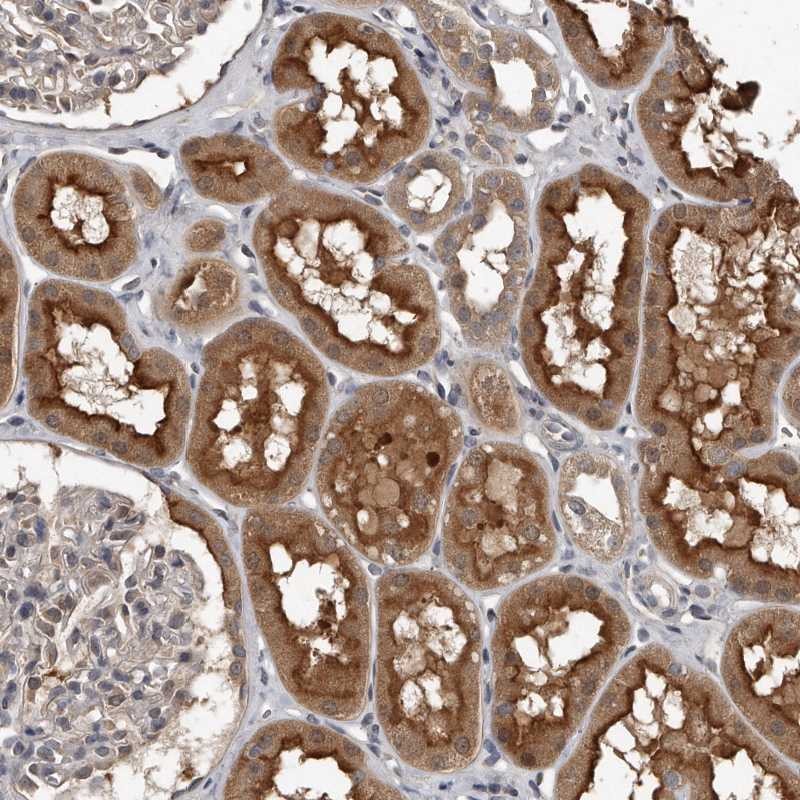
Immunohistochemical staining of human kidney shows strong cytoplasmic and membranous positivity in cells in tubules.
Immunohistochemical staining of human kidney shows strong cytoplasmic and membranous positivity in cells in tubules.
Anti-GRB10 Antibody
HPA027502
ApplicationsImmunoHistoChemistry
Product group Antibodies
ReactivityHuman
TargetGRB10
Overview
- SupplierAtlas Antibodies
- Product NameAnti-GRB10 Antibody
- Delivery Days Customer4
- ApplicationsImmunoHistoChemistry
- CertificationResearch Use Only
- ClonalityPolyclonal
- ConjugateUnconjugated
- Gene ID2887
- Target nameGRB10
- Target descriptiongrowth factor receptor bound protein 10
- Target synonymsGRB-IR, Grb-10, IRBP, MEG1, RSS, growth factor receptor-bound protein 10, GRB10 adapter protein, GRB10 adaptor protein, insulin receptor-binding protein Grb-IR, maternally expressed gene 1
- HostRabbit
- IsotypeIgG
- Protein IDQ13322
- Protein NameGrowth factor receptor-bound protein 10
- Scientific DescriptionRecombinant Protein Epitope Signature Tag (PrEST) antigen sequence
- ReactivityHuman
- Storage Instruction-20°C,2°C to 8°C
- UNSPSC41116161
Datasheet
MSDS
Related products
Product group Antibodies
GRB10 AntibodyCSB-PA002806
ApplicationsImmunoFluorescence, Western Blot, ELISA, ImmunoHistoChemistry
ReactivityHuman
TargetGRB10
- SizePrice

Product group Antibodies
Anti-GRB10 Antibody Picoband(r)A01663-2-CARRIER-FREE
ApplicationsFlow Cytometry, Western Blot, ELISA, ImmunoHistoChemistry
ReactivityHuman, Monkey, Mouse, Rat
TargetGRB10
- SizePrice

Product group Antibodies
Anti-GRB10 AntibodyA83162
ApplicationsWestern Blot, ELISA
ReactivityHuman
- SizePrice
Product group Antibodies

GRB10 AntibodyLS-C331035
ApplicationsWestern Blot
ReactivityHuman, Mouse
TargetGRB10
- SizePrice
Product group Antibodies

Goat anti-GRB10EB08476
ApplicationsWestern Blot, ELISA
ReactivityHuman
TargetGRB10
- SizePrice

Product group Antibodies
Anti-GRB10 AntibodyHPA031818
ApplicationsImmunoCytoChemistry
ReactivityHuman
TargetGRB10
- SizePrice
Product group Antibodies
ApplicationsWestern Blot, ImmunoHistoChemistry
ReactivityMouse
TargetGRB10
- SizePrice
Product group Antibodies
GRB10 Polyclonal AntibodyBS-2769R
ApplicationsImmunoFluorescence, Western Blot, ELISA, ImmunoCytoChemistry, ImmunoHistoChemistry, ImmunoHistoChemistry Frozen, ImmunoHistoChemistry Paraffin
ReactivityBovine, Canine, Equine, Human, Mouse, Porcine, Rabbit, Rat
TargetGRB10
- SizePrice