


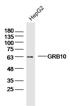
Figure 1. Western blot analysis of GRB10 using anti-GRB10 antibody (M01663). Electrophoresis was performed on a 5-20% SDS-PAGE gel at 70V (Stacking gel) / 90V (Resolving gel) for 2-3 hours. The sample well of each lane was loaded with 30ug of sample under reducing conditions. Lane 1: human Hek293 whole cell lysates, Lane 2: human Hela whole cell lysates, Lane 3: monkey COS-7 whole cell lysates, Lane 4: human HepG2 whole cell lysates, Lane 5: rat kidney tissue lysates, Lane 6: rat NRK whole cell lysates. After Electrophoresis, proteins were transferred to a Nitrocellulose membrane at 150mA for 50-90 minutes. Blocked the membrane with 5% Non-fat Milk/ TBS for 1.5 hour at RT. The membrane was incubated with mouse anti-GRB10 antigen affinity purified monoclonal antibody (Catalog # M01663) at 0.5 microg/mL overnight at 4°C, then washed with TBS-0.1%Tween 3 times with 5 minutes each and probed with a goat anti-mouse IgG-HRP secondary antibody at a dilution of 1:10000 for 1.5 hour at RT. The signal is developed using an Enhanced Chemiluminescent detection (ECL) kit (Catalog # EK1001) with Tanon 5200 system. A specific band was detected for GRB10 at approximately 67KD. The expected band size for GRB10 is at 67KD.
. Overlay histogram showing THP-1 cells stained with M01663 (Blue line). To facilitate intracellular staining, cells were fixed with 4% paraformaldehyde and permeabilized with permeabilization buffer. The cells were blocked with 10% normal goat serum. And then incubated with mouse anti- GRB10 Antibody (M01663, 1microg/1x106 cells) for 30 min at 20°C. DyLight®488 conjugated goat anti-mouse IgG (BA1126, 5-10microg/1x106 cells) was used as secondary antibody for 30 minutes at 20°C. Isotype control antibody (Green line) was mouse IgG (1microg/1x106) used under the same conditions. Unlabelled sample without incubation with primary antibody and secondary antibody (Red line) was used as a blank control.")
Figure 1. Western blot analysis of GRB10 using anti-GRB10 antibody (M01663). Electrophoresis was performed on a 5-20% SDS-PAGE gel at 70V (Stacking gel) / 90V (Resolving gel) for 2-3 hours. The sample well of each lane was loaded with 30ug of sample under reducing conditions. Lane 1: human Hek293 whole cell lysates, Lane 2: human Hela whole cell lysates, Lane 3: monkey COS-7 whole cell lysates, Lane 4: human HepG2 whole cell lysates, Lane 5: rat kidney tissue lysates, Lane 6: rat NRK whole cell lysates. After Electrophoresis, proteins were transferred to a Nitrocellulose membrane at 150mA for 50-90 minutes. Blocked the membrane with 5% Non-fat Milk/ TBS for 1.5 hour at RT. The membrane was incubated with mouse anti-GRB10 antigen affinity purified monoclonal antibody (Catalog # M01663) at 0.5 microg/mL overnight at 4°C, then washed with TBS-0.1%Tween 3 times with 5 minutes each and probed with a goat anti-mouse IgG-HRP secondary antibody at a dilution of 1:10000 for 1.5 hour at RT. The signal is developed using an Enhanced Chemiluminescent detection (ECL) kit (Catalog # EK1001) with Tanon 5200 system. A specific band was detected for GRB10 at approximately 67KD. The expected band size for GRB10 is at 67KD.
Anti-GRB10 Antibody Picoband(r) (monoclonal, 5H7)
M01663-DYLIGHT488
ApplicationsFlow Cytometry, Western Blot
Product group Antibodies
ReactivityHuman, Monkey, Rat
TargetGRB10
Overview
- SupplierBoster Bio
- Product NameAnti-GRB10 Antibody Picoband(r) (monoclonal, 5H7)
- Delivery Days Customer9
- Application Supplier NoteTested Species: In-house tested species with positive results. Other applications have not been tested. Optimal dilutions should be determined by end users.
- ApplicationsFlow Cytometry, Western Blot
- CertificationResearch Use Only
- ClonalityMonoclonal
- Clone ID5H7
- Concentration500 ug/ml
- ConjugateDyLight 488
- Gene ID2887
- Target nameGRB10
- Target descriptiongrowth factor receptor bound protein 10
- Target synonymsGRB-IR, Grb-10, IRBP, MEG1, RSS, growth factor receptor-bound protein 10, GRB10 adapter protein, GRB10 adaptor protein, insulin receptor-binding protein Grb-IR, maternally expressed gene 1
- HostMouse
- IsotypeIgG1
- Protein IDQ13322
- Protein NameGrowth factor receptor-bound protein 10
- Scientific DescriptionBoster Bio Anti-GRB10 Antibody Picoband® (monoclonal, 5H7) catalog # M01663. Tested in Flow Cytometry, WB applications. This antibody reacts with Human, Monkey, Rat. The brand Picoband indicates this is a premium antibody that guarantees superior quality, high affinity, and strong signals with minimal background in Western blot applications. Only our best-performing antibodies are designated as Picoband, ensuring unmatched performance.
- ReactivityHuman, Monkey, Rat
- Storage Instruction-20°C,2°C to 8°C
- UNSPSC12352203
Related products
Product group Antibodies
GRB10 Polyclonal AntibodyBS-2769R
ApplicationsImmunoFluorescence, Western Blot, ELISA, ImmunoCytoChemistry, ImmunoHistoChemistry, ImmunoHistoChemistry Frozen, ImmunoHistoChemistry Paraffin
ReactivityBovine, Canine, Equine, Human, Mouse, Porcine, Rabbit, Rat
TargetGRB10
- SizePrice
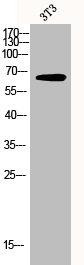
Product group Antibodies
ApplicationsWestern Blot, ImmunoHistoChemistry
ReactivityMouse
TargetGRB10
- SizePrice

Product group Antibodies
Anti-GRB10 AntibodyA83162
ApplicationsWestern Blot, ELISA
ReactivityHuman
- SizePrice
Product group Antibodies
Anti-GRB10 Antibody144-61629
ApplicationsWestern Blot
ReactivityHuman, Mouse
TargetGRB10
- SizePrice
Product group Antibodies

Goat anti-GRB10 AntibodyEB08476
ApplicationsWestern Blot, ELISA
ReactivityHuman
TargetGRB10
- SizePrice
Product group Antibodies
GRB10 antibodyGTX125980
ApplicationsWestern Blot, ImmunoHistoChemistry, ImmunoHistoChemistry Paraffin
ReactivityHuman
TargetGRB10
- SizePrice
Product group Antibodies

GRB10 AntibodyLS-C331035
ApplicationsWestern Blot
ReactivityHuman, Mouse
TargetGRB10
- SizePrice
Product group Antibodies
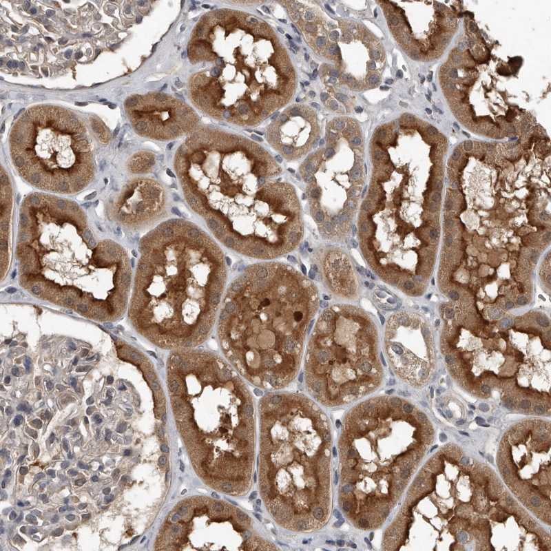
Anti-GRB10 AntibodyHPA027502
ApplicationsImmunoHistoChemistry
ReactivityHuman
TargetGRB10
- SizePrice
Product group Antibodies
GRB10 AntibodyCSB-PA002806
ApplicationsImmunoFluorescence, Western Blot, ELISA, ImmunoHistoChemistry
ReactivityHuman
TargetGRB10
- SizePrice