


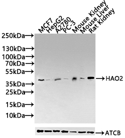
Figure 1. Western blot analysis of HAO2 using anti-HAO2 antibody (A11930-2). Electrophoresis was performed on a 5-20% SDS-PAGE gel at 70V (Stacking gel) / 90V (Resolving gel) for 2-3 hours. The sample well of each lane was loaded with 30 ug of sample under reducing conditions. Lane 1: human 293T whole cell lysates, Lane 2: human HepG2 whole cell lysates, Lane 3: rat liver tissue lysates, Lane 4: rat kidney tissue lysates, Lane 5: mouse liver tissue lysates, Lane 6: mouse kidney tissue lysates. After electrophoresis, proteins were transferred to a nitrocellulose membrane at 150 mA for 50-90 minutes. Blocked the membrane with 5% non-fat milk/TBS for 1.5 hour at RT. The membrane was incubated with rabbit anti-HAO2 antigen affinity purified polyclonal antibody (Catalog # A11930-2) at 0.5 microg/mL overnight at 4°C, then washed with TBS-0.1%Tween 3 times with 5 minutes each and probed with a goat anti-rabbit IgG-HRP secondary antibody at a dilution of 1:5000 for 1.5 hour at RT. The signal is developed using an Enhanced Chemiluminescent detection (ECL) kit (Catalog # EK1002) with Tanon 5200 system. A specific band was detected for HAO2 at approximately 39 kDa. The expected band size for HAO2 is at 39 kDa.
. Overlay histogram showing 293T cells stained with A11930-2 (Blue line). To facilitate intracellular staining, cells were fixed with 4% paraformaldehyde and permeabilized with permeabilization buffer. The cells were blocked with 10% normal goat serum. And then incubated with rabbit anti-HAO2 Antibody (A11930-2, 1 microg/1x106 cells) for 30 min at 20°C. DyLight®488 conjugated goat anti-rabbit IgG (BA1127, 5-10 microg/1x106 cells) was used as secondary antibody for 30 minutes at 20°C. Isotype control antibody (Green line) was rabbit IgG (1 microg/1x106) used under the same conditions. Unlabelled sample (Red line) was also used as a control.")
Figure 1. Western blot analysis of HAO2 using anti-HAO2 antibody (A11930-2). Electrophoresis was performed on a 5-20% SDS-PAGE gel at 70V (Stacking gel) / 90V (Resolving gel) for 2-3 hours. The sample well of each lane was loaded with 30 ug of sample under reducing conditions. Lane 1: human 293T whole cell lysates, Lane 2: human HepG2 whole cell lysates, Lane 3: rat liver tissue lysates, Lane 4: rat kidney tissue lysates, Lane 5: mouse liver tissue lysates, Lane 6: mouse kidney tissue lysates. After electrophoresis, proteins were transferred to a nitrocellulose membrane at 150 mA for 50-90 minutes. Blocked the membrane with 5% non-fat milk/TBS for 1.5 hour at RT. The membrane was incubated with rabbit anti-HAO2 antigen affinity purified polyclonal antibody (Catalog # A11930-2) at 0.5 microg/mL overnight at 4°C, then washed with TBS-0.1%Tween 3 times with 5 minutes each and probed with a goat anti-rabbit IgG-HRP secondary antibody at a dilution of 1:5000 for 1.5 hour at RT. The signal is developed using an Enhanced Chemiluminescent detection (ECL) kit (Catalog # EK1002) with Tanon 5200 system. A specific band was detected for HAO2 at approximately 39 kDa. The expected band size for HAO2 is at 39 kDa.
Anti-HAO2 Antibody Picoband(r)
A11930-2-PE
ApplicationsFlow Cytometry, Western Blot, ELISA
Product group Antibodies
ReactivityHuman, Mouse, Rat
TargetHAO2
Overview
- SupplierBoster Bio
- Product NameAnti-HAO2 Antibody Picoband(r)
- Delivery Days Customer9
- ApplicationsFlow Cytometry, Western Blot, ELISA
- CertificationResearch Use Only
- ClonalityPolyclonal
- Concentration500 ug/ml
- ConjugateRPE
- Gene ID51179
- Target nameHAO2
- Target descriptionhydroxyacid oxidase 2
- Target synonymsGIG16, HAOX2, 2-Hydroxyacid oxidase 2, (S)-2-hydroxy-acid oxidase, peroxisomal, cell growth-inhibiting gene 16 protein, glycolate oxidase, hydroxyacid oxidase 2 (long chain), long chain alpha-hydroxy acid oxidase, long-chain L-2-hydroxy acid oxidase
- HostRabbit
- IsotypeIgG
- Protein IDQ9NYQ3
- Protein Name2-Hydroxyacid oxidase 2
- Scientific DescriptionBoster Bio Anti-HAO2 Antibody Picoband® catalog # A11930-2. Tested in ELISA, WB, Flow Cytometry applications. This antibody reacts with Human, Mouse, Rat. The brand Picoband indicates this is a premium antibody that guarantees superior quality, high affinity, and strong signals with minimal background in Western blot applications. Only our best-performing antibodies are designated as Picoband, ensuring unmatched performance.
- ReactivityHuman, Mouse, Rat
- Storage Instruction-20°C,2°C to 8°C
- UNSPSC12352203
Related products
Product group Antibodies
HAO2 AntibodyCSB-PA882145LA01HU
ApplicationsWestern Blot, ELISA, ImmunoHistoChemistry
ReactivityHuman, Mouse, Rat
TargetHAO2
- SizePrice
Product group Antibodies

ApplicationsWestern Blot, ELISA
ReactivityHuman
TargetHAO2
- SizePrice
Product group Antibodies
Anti-HAO2 Antibody Picoband(r)A11930-2-CARRIER-FREE
ApplicationsFlow Cytometry, Western Blot, ELISA
ReactivityHuman, Mouse, Rat
TargetHAO2
- SizePrice
Product group Antibodies
Hao2 Polyclonal AntibodyCAC08083
ApplicationsWestern Blot, ELISA, ImmunoHistoChemistry
ReactivityMouse, Rat
TargetHAO2
- SizePrice
Product group Antibodies
HAO2 antibody, N-termGTX47157
ApplicationsWestern Blot
ReactivityHuman
TargetHAO2
- SizePrice
Product group Antibodies

HAO2 AntibodyLS-C750133
ApplicationsWestern Blot
ReactivityHuman, Mouse
TargetHAO2
- SizePrice
![Lane 1: Marker [kDa] 250, 130, 95, 72, 55, 36, 28, 17, 10 | Lane 2: RT4 | Lane 3: U-251 MG | Lane 4: Human Plasma | Lane 5: Liver | Lane 6: Tonsil](https://atlasantibodies.s3.amazonaws.com/images/wb/hpa050355-wb-1.jpg)
Product group Antibodies
Anti-HAO2 AntibodyHPA050355
ApplicationsWestern Blot
ReactivityHuman
TargetHAO2
- SizePrice