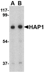
Bio-Connect
Anti-HAP1 Antibody

Anti-HAP1 Antibody

A48372
Antibodies.com
ApplicationsWestern Blot, ELISA, ImmunoHistoChemistry
Product group Antibodies
Sign in to order and to see your custom pricing.
Large volume orders?
Order with a bulk request

Overview

  • Supplier
    Antibodies.com
  • Product Name
    Anti-HAP1 Antibody
  • Delivery Days Customer
    7
  • Applications
    Western Blot, ELISA, ImmunoHistoChemistry
  • Certification
    Research Use Only
  • Clonality
    Polyclonal
  • Conjugate
    Unconjugated
  • Host
    Rabbit
  • Scientific Description
    Rabbit polyclonal antibody to HAP1
  • UNSPSC
    12352203

Related products

Product group Antibodies
Biomatik
ApplicationsImmunoPrecipitation, Western Blot, ImmunoCytoChemistry, ImmunoHistoChemistry
TargetHAP1
  • SizePrice
Immunofluorescence staining of SH-SY5Y cells with CSB-PA010129LA01HU at 1:100, counter-stained with DAPI. The cells were fixed in 4% formaldehyde, permeabilized using 0.2% Triton X-100 and blocked in 10% normal Goat Serum. The cells were then incubated with the antibody overnight at 4°C. The secondary antibody was Alexa Fluor 488-congugated AffiniPure Goat Anti-Rabbit IgG(H+L).
Product group Antibodies
Cusabio
HAP1 AntibodyCSB-PA010129LA01HU
ApplicationsImmunoFluorescence, ELISA
ReactivityHuman
TargetHAP1
  • SizePrice
Product group Antibodies
LifeSpan BioSciences
ApplicationsWestern Blot
TargetHAP1
  • SizePrice
Figure 1. Western blot analysis of HAP1 using anti-HAP1 antibody (A01658-3). Electrophoresis was performed on a 5-20% SDS-PAGE gel at 70V (Stacking gel) / 90V (Resolving gel) for 2-3 hours. The sample well of each lane was loaded with 30ug of sample under reducing conditions. Lane 1: rat liver tissue lysates, Lane 2: mouse liver tissue lysates, Lane 3: mouse Neuro-2a whole cell lysates. After Electrophoresis, proteins were transferred to a Nitrocellulose membrane at 150mA for 50-90 minutes. Blocked the membrane with 5% Non-fat Milk/ TBS for 1.5 hour at RT. The membrane was incubated with rabbit anti-HAP1 antigen affinity purified polyclonal antibody (Catalog # A01658-3) at 0.5 microg/mL overnight at 4°C, then washed with TBS-0.1%Tween 3 times with 5 minutes each and probed with a goat anti-rabbit IgG-HRP secondary antibody at a dilution of 1:5000 for 1.5 hour at RT. The signal is developed using an Enhanced Chemiluminescent detection (ECL) kit (Catalog # EK1002) with Tanon 5200 system. A specific band was detected for HAP1 at approximately 80KD. The expected band size for HAP1 is at 80KD.
Product group Antibodies
Boster Bio
Anti-HAP1 Antibody Picoband(r)A01658-3-CARRIER-FREE
ApplicationsFlow Cytometry, ImmunoFluorescence, Western Blot, ELISA, ImmunoCytoChemistry
TargetHAP1
  • SizePrice
Product group Antibodies
Everest Biotech
ApplicationsWestern Blot, ELISA, ImmunoHistoChemistry
TargetHAP1
  • SizePrice
WB analysis of human brain (hippocampus) lysate using GTX89202 HAP1 antibody, Internal. Dilution : 0.1microg/ml Loading : 35microg protein in RIPA buffer
Product group Antibodies
GeneTex
ApplicationsWestern Blot, ImmunoHistoChemistry, ImmunoHistoChemistry Paraffin
TargetHAP1
  • SizePrice