

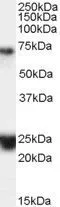
WB analysis of human brain (hippocampus) lysate using GTX89202 HAP1 antibody, Internal. Dilution : 0.1μg/ml Loading : 35μg protein in RIPA buffer

WB analysis of human brain (hippocampus) lysate using GTX89202 HAP1 antibody, Internal. Dilution : 0.1μg/ml Loading : 35μg protein in RIPA buffer
HAP1 antibody, Internal
GTX89202
ApplicationsWestern Blot, ImmunoHistoChemistry, ImmunoHistoChemistry Paraffin
Product group Antibodies
ReactivityHuman
TargetHAP1
Overview
- SupplierGeneTex
- Product NameHAP1 antibody, Internal
- Delivery Days Customer7
- Application Supplier NoteWB: 0.1-0.3microg/ml. IHC-P: 3.75microg/ml. *Optimal dilutions/concentrations should be determined by the researcher.Not tested in other applications.
- ApplicationsWestern Blot, ImmunoHistoChemistry, ImmunoHistoChemistry Paraffin
- CertificationResearch Use Only
- ClonalityPolyclonal
- Concentration0.50 mg/ml
- ConjugateUnconjugated
- Gene ID9001
- Target nameHAP1
- Target descriptionhuntingtin associated protein 1
- Target synonymsHAP2, HIP5, HLP, hHLP1, huntingtin-associated protein 1, HAP-1, epididymis secretory sperm binding protein, huntingtin-associated protein 2, neuroan 1
- HostGoat
- IsotypeIgG
- Protein IDP54257
- Protein NameHuntingtin-associated protein 1
- Scientific DescriptionHuntingtons disease (HD), a neurodegenerative disorder characterized by loss of striatal neurons, is caused by an expansion of a polyglutamine tract in the HD protein huntingtin. This gene encodes a protein that interacts with huntingtin, with two cytoskeletal proteins (dynactin and pericentriolar autoantigen protein 1), and with a hepatocyte growth factor-regulated tyrosine kinase substrate. The interactions with cytoskeletal proteins and a kinase substrate suggest a role for this protein in vesicular trafficking or organelle transport. Several alternatively spliced transcript variants encoding different isoforms have been described for this gene. [provided by RefSeq, Jul 2008]
- ReactivityHuman
- Storage Instruction-20°C or -80°C,2°C to 8°C
- UNSPSC41116161
Datasheet
Related products

Product group Antibodies
Anti-HAP1 Antibody Picoband(r)A01658-3-CARRIER-FREE
ApplicationsFlow Cytometry, ImmunoFluorescence, Western Blot, ELISA, ImmunoCytoChemistry
ReactivityHuman, Mouse, Rat
TargetHAP1
- SizePrice
Product group Antibodies
Anti-HAP1 AntibodyA48372
ApplicationsWestern Blot, ELISA, ImmunoHistoChemistry
ReactivityHuman, Mouse, Rat
- SizePrice
Product group Antibodies

Goat anti-HAP1EB07528
ApplicationsWestern Blot, ELISA, ImmunoHistoChemistry
ReactivityHuman
TargetHAP1
- SizePrice
Product group Antibodies
HAP1 AntibodyCSB-PA010129LA01HU
ApplicationsImmunoFluorescence, ELISA
ReactivityHuman
TargetHAP1
- SizePrice
Product group Antibodies

HAP1 Antibody (aa160-381)LS-C373623
ApplicationsWestern Blot
ReactivityHuman
TargetHAP1
- SizePrice
Product group Antibodies
ApplicationsImmunoPrecipitation, Western Blot, ImmunoCytoChemistry, ImmunoHistoChemistry
ReactivityPorcine
TargetHAP1
- SizePrice