


Anti-HAS1 Antibody
A11318
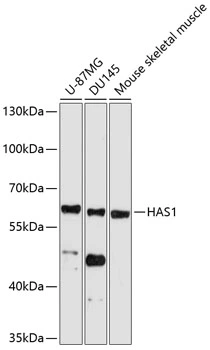
ApplicationsWestern Blot
Product group Antibodies
ReactivityHuman, Mouse
Overview
- SupplierAntibodies.com
- Product NameAnti-HAS1 Antibody
- Delivery Days Customer7
- ApplicationsWestern Blot
- CertificationResearch Use Only
- ClonalityPolyclonal
- ConjugateUnconjugated
- HostRabbit
- IsotypeIgG
- Scientific DescriptionRabbit polyclonal antibody to HAS1.
- ReactivityHuman, Mouse
- UNSPSC12352203
Related products

Product group Antibodies
Anti-Hyaluronan synthase 1/HAS1 Antibody Picoband(r)A04784-1-CARRIER-FREE
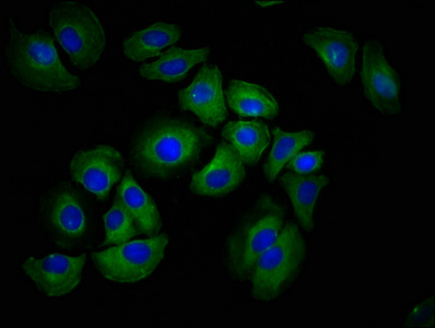
ApplicationsImmunoFluorescence, Western Blot, ImmunoCytoChemistry, ImmunoHistoChemistry
ReactivityHuman, Mouse, Rat
TargetHAS1
- SizePrice
Product group Antibodies
Anti-HAS1 Antibody144-61887
ApplicationsWestern Blot
ReactivityHuman, Mouse
TargetHAS1
- SizePrice
Product group Antibodies

HAS1 / HAS AntibodyLS-C831068
ApplicationsELISA, ImmunoHistoChemistry
ReactivityHuman, Mouse
TargetHAS1
- SizePrice
Product group Antibodies
HAS1 AntibodyCSB-PA010139LA01HU
ApplicationsImmunoFluorescence, ELISA
ReactivityHuman
TargetHAS1
- SizePrice
Product group Antibodies
References
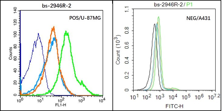
HAS1 Polyclonal AntibodyBS-2946R
ApplicationsFlow Cytometry, ImmunoFluorescence, ELISA, ImmunoCytoChemistry, ImmunoHistoChemistry, ImmunoHistoChemistry Frozen, ImmunoHistoChemistry Paraffin
ReactivityBovine, Human, Mouse, Porcine, Rat, Sheep
TargetHAS1
- SizePrice
Product group Antibodies
HAS1 antibodyGTX04887
ApplicationsWestern Blot
ReactivityHuman, Mouse
TargetHAS1
- SizePrice