

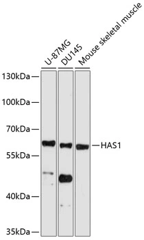
WB analysis of various samples using HAS1 antibody. Loading : 25 μg Dilution : 1:1000
WB analysis of various samples using HAS1 antibody. Loading : 25 μg Dilution : 1:1000
HAS1 antibody
GTX04887
ApplicationsWestern Blot
Product group Antibodies
ReactivityHuman, Mouse
TargetHAS1
Overview
- SupplierGeneTex
- Product NameHAS1 antibody
- Delivery Days Customer7
- Application Supplier NoteWB: 1:500-1:2000. *Optimal dilutions/concentrations should be determined by the researcher.Not tested in other applications.
- ApplicationsWestern Blot
- CertificationResearch Use Only
- ClonalityPolyclonal
- ConjugateUnconjugated
- Gene ID3036
- Target nameHAS1
- Target descriptionhyaluronan synthase 1
- Target synonymsHAS, hyaluronan synthase 1, HA synthase 1, hyaluronate synthase 1, hyaluronic acid synthase 1
- HostRabbit
- IsotypeIgG
- Protein IDQ92839
- Protein NameHyaluronan synthase 1
- Scientific DescriptionHyaluronan or hyaluronic acid (HA) is a high molecular weight unbranched polysaccharide synthesized by a wide variety of organisms from bacteria to mammals, and is a constituent of the extracellular matrix. It consists of alternating glucuronic acid and N-acetylglucosamine residues that are linked by beta-1-3 and beta-1-4 glycosidic bonds. HA is synthesized by membrane-bound synthase at the inner surface of the plasma membrane, and the chains are extruded through pore-like structures into the extracellular space. It serves a variety of functions, including space filling, lubrication of joints, and provision of a matrix through which cells can migrate. HA is actively produced during wound healing and tissue repair to provide a framework for ingrowth of blood vessels and fibroblasts. Changes in the serum concentration of HA are associated with inflammatory and degenerative arthropathies such as rheumatoid arthritis. In addition, the interaction of HA with the leukocyte receptor CD44 is important in tissue-specific homing by leukocytes, and overexpression of HA receptors has been correlated with tumor metastasis. HAS1 is a member of the newly identified vertebrate gene family encoding putative hyaluronan synthases, and its amino acid sequence shows significant homology to the hasA gene product of Streptococcus pyogenes, a glycosaminoglycan synthetase (DG42) from Xenopus laevis, and a recently described murine hyaluronan synthase. Alternative splicing results in multiple transcript variants. [provided by RefSeq, Jul 2014]
- ReactivityHuman, Mouse
- Storage Instruction-20°C or -80°C,2°C to 8°C
- UNSPSC41116161
Datasheet
Related products

Product group Antibodies
Anti-Hyaluronan synthase 1/HAS1 Antibody Picoband(r)A04784-1-CARRIER-FREE
ApplicationsImmunoFluorescence, Western Blot, ImmunoCytoChemistry, ImmunoHistoChemistry
ReactivityHuman, Mouse, Rat
TargetHAS1
- SizePrice

Product group Antibodies
Anti-HAS1 AntibodyA11318
ApplicationsWestern Blot
ReactivityHuman, Mouse
- SizePrice
Product group Antibodies
Anti-HAS1 Antibody144-61887
ApplicationsWestern Blot
ReactivityHuman, Mouse
TargetHAS1
- SizePrice
Product group Antibodies

HAS1 / HAS AntibodyLS-C831068
ApplicationsELISA, ImmunoHistoChemistry
ReactivityHuman, Mouse
TargetHAS1
- SizePrice
Product group Antibodies
HAS1 AntibodyCSB-PA010139LA01HU
ApplicationsImmunoFluorescence, ELISA
ReactivityHuman
TargetHAS1
- SizePrice
Product group Antibodies
References
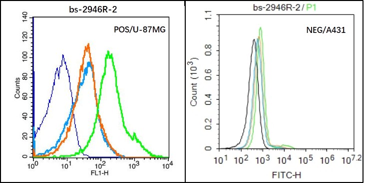
HAS1 Polyclonal AntibodyBS-2946R
ApplicationsFlow Cytometry, ImmunoFluorescence, ELISA, ImmunoCytoChemistry, ImmunoHistoChemistry, ImmunoHistoChemistry Frozen, ImmunoHistoChemistry Paraffin
ReactivityBovine, Human, Mouse, Porcine, Rat, Sheep
TargetHAS1
- SizePrice
![WB analysis of human HAS1 (AA: 74-243) recombinant protein using GTX82799 HAS1 antibody [3E10].](https://www.genetex.com/upload/website/prouct_img/normal/GTX82799/GTX82799_20170912_WB_w_23061322_124.webp)
Product group Antibodies
References
HAS1 antibody [3E10]GTX82799
ApplicationsImmunoFluorescence, Western Blot, ELISA, ImmunoCytoChemistry, ImmunoHistoChemistry, ImmunoHistoChemistry Paraffin
ReactivityHuman
TargetHAS1
- SizePrice
Product group Antibodies
HAS1 antibodyGTX60186
ApplicationsImmunoHistoChemistry, ImmunoHistoChemistry Paraffin
ReactivityHuman, Mouse, Rat
TargetHAS1
- SizePrice