

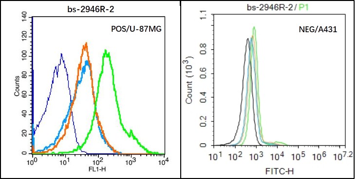
U-87MG (Positive) and A431 (Negative control) cells (black) were incubated in 5% BSA blocking buffer for 30 min at room temperature. Cells were then stained with HAS1 Antibody (bs-2946R) at 1:50 dilution in blocking buffer and incubated for 30 min at room temperature, washed twice with 2% BSA in PBS, followed by secondary antibody(blue) incubation for 40 min at room temperature. Acquisitions of 20,000 events were performed. Cells stained with primary antibody (green), and isotype control (orange)._x000D_
 at 1:200 followed by conjugation to the secondary antibody and DAB staining\n")
U-87MG (Positive) and A431 (Negative control) cells (black) were incubated in 5% BSA blocking buffer for 30 min at room temperature. Cells were then stained with HAS1 Antibody (bs-2946R) at 1:50 dilution in blocking buffer and incubated for 30 min at room temperature, washed twice with 2% BSA in PBS, followed by secondary antibody(blue) incubation for 40 min at room temperature. Acquisitions of 20,000 events were performed. Cells stained with primary antibody (green), and isotype control (orange)._x000D_
HAS1 Polyclonal Antibody
BS-2946R
ApplicationsFlow Cytometry, ImmunoFluorescence, ELISA, ImmunoCytoChemistry, ImmunoHistoChemistry, ImmunoHistoChemistry Frozen, ImmunoHistoChemistry Paraffin
Product group Antibodies
ReactivityBovine, Human, Mouse, Porcine, Rat, Sheep
TargetHAS1
Overview
- SupplierBioss
- Product NameHAS1 Polyclonal Antibody
- Delivery Days Customer16
- ApplicationsFlow Cytometry, ImmunoFluorescence, ELISA, ImmunoCytoChemistry, ImmunoHistoChemistry, ImmunoHistoChemistry Frozen, ImmunoHistoChemistry Paraffin
- Applications SupplierELISA(1:500-1000), FCM(1:20-100), IHC-P(1:200-400), IHC-F(1:100-500), IF(IHC-P)(1:50-200), IF(IHC-F)(1:50-200), IF(ICC)(1:50-200)
- CertificationResearch Use Only
- ClonalityPolyclonal
- Concentration1 ug/ul
- ConjugateUnconjugated
- Gene ID3036
- Target nameHAS1
- Target descriptionhyaluronan synthase 1
- Target synonymsHAS, hyaluronan synthase 1, HA synthase 1, hyaluronate synthase 1, hyaluronic acid synthase 1
- HostRabbit
- IsotypeIgG
- Protein IDQ92839
- Protein NameHyaluronan synthase 1
- ReactivityBovine, Human, Mouse, Porcine, Rat, Sheep
- Storage Instruction-20°C
- UNSPSC41116161
References
- Hyaluronan and hyaluronan synthases expression and localization in embryonic mouse molars. Yang G et al., 2016 Aug, J Mol HistolRead this paper
Datasheet
Related products

Product group Antibodies
Anti-Hyaluronan synthase 1/HAS1 Antibody Picoband(r)A04784-1-CARRIER-FREE
ApplicationsImmunoFluorescence, Western Blot, ImmunoCytoChemistry, ImmunoHistoChemistry
ReactivityHuman, Mouse, Rat
TargetHAS1
- SizePrice

Product group Antibodies
Anti-HAS1 AntibodyA11318
ApplicationsWestern Blot
ReactivityHuman, Mouse
- SizePrice
Product group Antibodies
Anti-HAS1 Antibody144-61887
ApplicationsWestern Blot
ReactivityHuman, Mouse
TargetHAS1
- SizePrice
Product group Antibodies

HAS1 / HAS AntibodyLS-C831068
ApplicationsELISA, ImmunoHistoChemistry
ReactivityHuman, Mouse
TargetHAS1
- SizePrice
Product group Antibodies
HAS1 AntibodyCSB-PA010139LA01HU
ApplicationsImmunoFluorescence, ELISA
ReactivityHuman
TargetHAS1
- SizePrice
Product group Antibodies
HAS1 antibodyGTX04887
ApplicationsWestern Blot
ReactivityHuman, Mouse
TargetHAS1
- SizePrice