


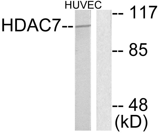
Figure 1. Western blot analysis of HDAC7 using anti-HDAC7 antibody (PB9629). Electrophoresis was performed on a 5-20% SDS-PAGE gel at 70V (Stacking gel) / 90V (Resolving gel) for 2-3 hours. Lane 1: Rat Spleen Tissue Lysate at 50ug, Lane 2: Rat Thymus Tissue Lysate at 50ug, Lane 3: Human Placenta Tissue Lysate at 50ug, Lane 4: HELA Whole Cell Lysate at 40ug. After electrophoresis, proteins were transferred to a nitrocellulose membrane at 150 mA for 50-90 minutes. Blocked the membrane with 5% non-fat milk/TBS for 1.5 hour at RT. The membrane was incubated with rabbit anti-HDAC7 antigen affinity purified polyclonal antibody (Catalog # PB9629) at 0.5 microg/mL overnight at 4°C, then washed with TBS-0.1%Tween 3 times with 5 minutes each and probed with a goat anti-rabbit IgG-HRP secondary antibody at a dilution of 1:5000 for 1.5 hour at RT. The signal is developed using an Enhanced Chemiluminescent detection (ECL) kit (Catalog # EK1002) with Tanon 5200 system. A specific band was detected for HDAC7 at approximately 103 kDa. The expected band size for HDAC7 is at 103 kDa.
Figure 1. Western blot analysis of HDAC7 using anti-HDAC7 antibody (PB9629). Electrophoresis was performed on a 5-20% SDS-PAGE gel at 70V (Stacking gel) / 90V (Resolving gel) for 2-3 hours. Lane 1: Rat Spleen Tissue Lysate at 50ug, Lane 2: Rat Thymus Tissue Lysate at 50ug, Lane 3: Human Placenta Tissue Lysate at 50ug, Lane 4: HELA Whole Cell Lysate at 40ug. After electrophoresis, proteins were transferred to a nitrocellulose membrane at 150 mA for 50-90 minutes. Blocked the membrane with 5% non-fat milk/TBS for 1.5 hour at RT. The membrane was incubated with rabbit anti-HDAC7 antigen affinity purified polyclonal antibody (Catalog # PB9629) at 0.5 microg/mL overnight at 4°C, then washed with TBS-0.1%Tween 3 times with 5 minutes each and probed with a goat anti-rabbit IgG-HRP secondary antibody at a dilution of 1:5000 for 1.5 hour at RT. The signal is developed using an Enhanced Chemiluminescent detection (ECL) kit (Catalog # EK1002) with Tanon 5200 system. A specific band was detected for HDAC7 at approximately 103 kDa. The expected band size for HDAC7 is at 103 kDa.
Anti-HDAC7 Antibody Picoband(r)
PB9629-CARRIER-FREE
ApplicationsWestern Blot
Product group Antibodies
ReactivityHuman, Rat
TargetHDAC7
Overview
- SupplierBoster Bio
- Product NameAnti-HDAC7 Antibody Picoband(r)
- Delivery Days Customer9
- Application Supplier NoteTested Species: In-house tested species with positive results. Other applications have not been tested. Optimal dilutions should be determined by end users.
- ApplicationsWestern Blot
- CertificationResearch Use Only
- ClonalityPolyclonal
- Concentration500 ug/ml
- Gene ID51564
- Target nameHDAC7
- Target descriptionhistone deacetylase 7
- Target synonymsHD7, HD7A, HDAC7A, histone deacetylase 7, histone deacetylase 7A, protein deacetylase HDAC7
- HostRabbit
- IsotypeIgG
- Protein IDQ8WUI4
- Protein NameHistone deacetylase 7
- Scientific DescriptionBoster Bio Anti-HDAC7 Antibody Picoband® catalog # PB9629. Tested in WB applications. This antibody reacts with Human, Rat. The brand Picoband indicates this is a premium antibody that guarantees superior quality, high affinity, and strong signals with minimal background in Western blot applications. Only our best-performing antibodies are designated as Picoband, ensuring unmatched performance.
- ReactivityHuman, Rat
- Storage Instruction-20°C,2°C to 8°C
- UNSPSC12352203
Related products
Product group Antibodies
HDAC7 AntibodyCSB-PA002897
ApplicationsWestern Blot, ELISA
ReactivityHuman, Mouse, Rat
TargetHDAC7
- SizePrice
Product group Antibodies
Anti-HDAC7 AntibodyA97489
ApplicationsWestern Blot, ELISA, ImmunoHistoChemistry
ReactivityHuman, Mouse
- SizePrice
Product group Antibodies

HDAC7A (Phospho-Ser155) AntibodyABX012724
ApplicationsWestern Blot, ELISA
- SizePrice
Product group Antibodies

Anti-HDAC7 [RAB-C321]Ab01782-1.1
ApplicationsImmunoFluorescence, ImmunoPrecipitation
ReactivityHuman
TargetHDAC7
- SizePrice
Product group Antibodies

HDAC7 Antibody (N-Terminus)LS-C359012
ApplicationsImmunoFluorescence, Western Blot, ImmunoCytoChemistry
ReactivityHuman, Mouse, Rat, Zebra Fish
TargetHDAC7
- SizePrice
Product group Antibodies
HDAC7 antibody [C2C3], C-termGTX114179
ApplicationsImmunoFluorescence, ImmunoPrecipitation, Western Blot, ImmunoCytoChemistry, ImmunoHistoChemistry, ImmunoHistoChemistry Paraffin
ReactivityHuman
TargetHDAC7
- SizePrice
Product group Antibodies
ApplicationsImmunoFluorescence, Western Blot, ELISA, ImmunoCytoChemistry, ImmunoHistoChemistry, ImmunoHistoChemistry Frozen, ImmunoHistoChemistry Paraffin
ReactivityBovine, Canine, Chicken, Equine, Human, Mouse, Rabbit, Rat
TargetHDAC7
- SizePrice
Product group Antibodies
Anti-HDAC7 Antibody144-07285
ApplicationsImmunoFluorescence, Western Blot
ReactivityHuman, Mouse, Rat
TargetHDAC7
- SizePrice