

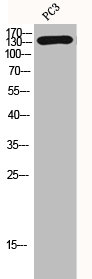
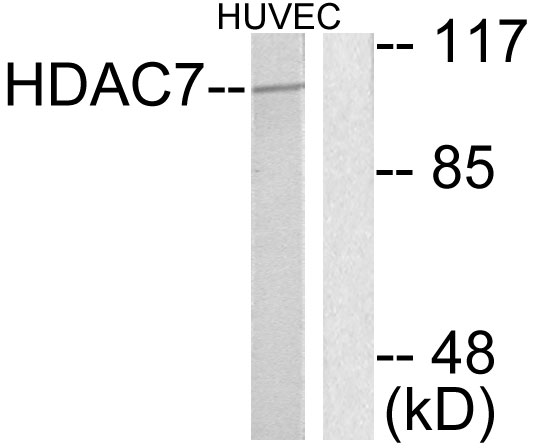
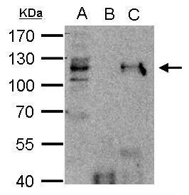
HDAC7A (Phospho-Ser155) Antibody
ABX012724
ApplicationsWestern Blot, ELISA
Product group Antibodies
Overview
- SupplierAbbexa
- Product NameHDAC7A (Phospho-Ser155) Antibody
- Delivery Days Customer12
- ApplicationsWestern Blot, ELISA
- CertificationResearch Use Only
- ClonalityPolyclonal
- ConjugateUnconjugated
- HostRabbit
- UNSPSC12352203
Related products
Product group Antibodies
HDAC7 AntibodyCSB-PA002897
ApplicationsWestern Blot, ELISA
ReactivityHuman, Mouse, Rat
TargetHDAC7
- SizePrice
Product group Antibodies
Anti-HDAC7 AntibodyA97489
ApplicationsWestern Blot, ELISA, ImmunoHistoChemistry
ReactivityHuman, Mouse
- SizePrice
Product group Antibodies

Anti-HDAC7 [RAB-C321]Ab01782-1.1
ApplicationsImmunoFluorescence, ImmunoPrecipitation
ReactivityHuman
TargetHDAC7
- SizePrice
Product group Antibodies

HDAC7 Antibody (N-Terminus)LS-C359012
ApplicationsImmunoFluorescence, Western Blot, ImmunoCytoChemistry
ReactivityHuman, Mouse, Rat, Zebra Fish
TargetHDAC7
- SizePrice

Product group Antibodies
Anti-HDAC7 Antibody Picoband(r)PB9629-CARRIER-FREE
ApplicationsWestern Blot
ReactivityHuman, Rat
TargetHDAC7
- SizePrice
Product group Antibodies
HDAC7 antibody [C2C3], C-termGTX114179
ApplicationsImmunoFluorescence, ImmunoPrecipitation, Western Blot, ImmunoCytoChemistry, ImmunoHistoChemistry, ImmunoHistoChemistry Paraffin
ReactivityHuman
TargetHDAC7
- SizePrice
Product group Antibodies
ApplicationsImmunoFluorescence, Western Blot, ELISA, ImmunoCytoChemistry, ImmunoHistoChemistry, ImmunoHistoChemistry Frozen, ImmunoHistoChemistry Paraffin
ReactivityBovine, Canine, Chicken, Equine, Human, Mouse, Rabbit, Rat
TargetHDAC7
- SizePrice
Product group Antibodies
Anti-HDAC7 Antibody144-07285
ApplicationsImmunoFluorescence, Western Blot
ReactivityHuman, Mouse, Rat
TargetHDAC7
- SizePrice