


Anti-HDAC9 Antibody
A98657
ApplicationsImmunoFluorescence, Western Blot, ELISA, ImmunoHistoChemistry
Product group Antibodies
ReactivityHuman
Overview
- SupplierAntibodies.com
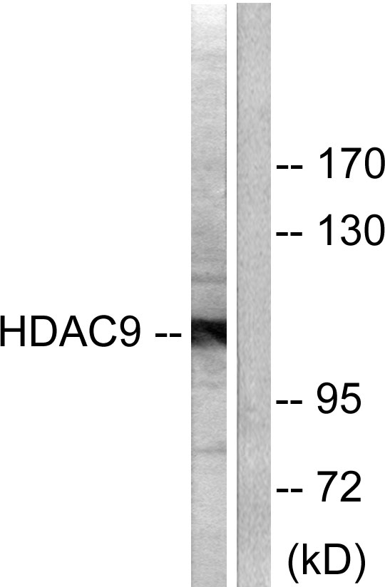
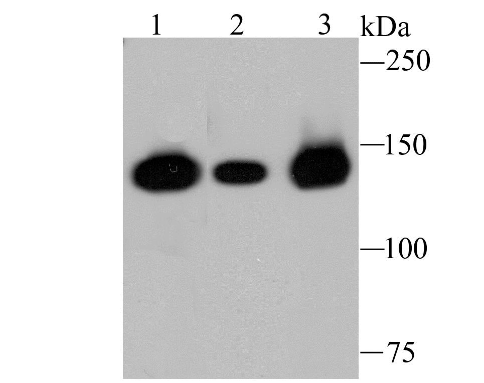
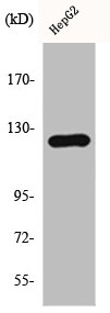
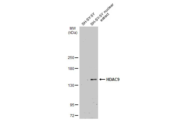
- Product NameAnti-HDAC9 Antibody
- Delivery Days Customer7
- ApplicationsImmunoFluorescence, Western Blot, ELISA, ImmunoHistoChemistry
- CertificationResearch Use Only
- ClonalityPolyclonal
- ConjugateUnconjugated
- HostRabbit
- IsotypeIgG
- Scientific DescriptionRabbit polyclonal antibody to HDAC9.
- ReactivityHuman
- UNSPSC12352203
Related products
Product group Antibodies
Anti-HDAC9 Antibody144-01516
ApplicationsWestern Blot
ReactivityHuman, Mouse, Rat
TargetHDAC9
- SizePrice


Product group Antibodies
Anti-HDAC9 Antibody Picoband(r)A02177-4-CARRIER-FREE
ApplicationsFlow Cytometry, ImmunoFluorescence, Western Blot, ELISA, ImmunoCytoChemistry
ReactivityHuman, Mouse, Rat
TargetHDAC9
- SizePrice
Product group Antibodies
HDAC9 Recombinant AntibodyBSM-54186R
ApplicationsImmunoFluorescence, Western Blot, ImmunoCytoChemistry, ImmunoHistoChemistry, ImmunoHistoChemistry Frozen, ImmunoHistoChemistry Paraffin
ReactivityHuman
TargetHDAC9
- SizePrice
Product group Antibodies
HDAC5/HDAC9 AntibodyCSB-PA002890
ApplicationsWestern Blot, ELISA, ImmunoHistoChemistry
ReactivityHuman, Mouse
TargetHDAC9
- SizePrice
Product group Antibodies
ApplicationsImmunoPrecipitation, Western Blot, ImmunoCytoChemistry, ImmunoHistoChemistry
ReactivityRat
TargetHDAC9
- SizePrice
Product group Antibodies
HDAC9 antibodyGTX132949
ApplicationsWestern Blot, ImmunoHistoChemistry, ImmunoHistoChemistry Paraffin
ReactivityHuman, Mouse
TargetHDAC9
- SizePrice
Product group Antibodies

HDAC9 AntibodyLS-C331507
ApplicationsWestern Blot
ReactivityHuman, Mouse, Rat
TargetHDAC9
- SizePrice

Product group Antibodies
Anti-HDAC9 AntibodyHPA028926
ApplicationsImmunoCytoChemistry, ImmunoHistoChemistry
ReactivityHuman
TargetHDAC9
- SizePrice