


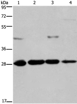
Figure 1. Western blot analysis of Hes1 using anti-Hes1 antibody (M01459). Electrophoresis was performed on a 5-20% SDS-PAGE gel at 70V (Stacking gel) / 90V (Resolving gel) for 2-3 hours. The sample well of each lane was loaded with 30 ug of sample under reducing conditions. Lane 1: human U-87MG whole cell lysates, Lane 2: human U251 whole cell lysates, Lane 3: human 293T whole cell lysates, Lane 4: rat brain tissue lysates, Lane 5: mouse brain tissue lysates. After electrophoresis, proteins were transferred to a nitrocellulose membrane at 150 mA for 50-90 minutes. Blocked the membrane with 5% non-fat milk/TBS for 1.5 hour at RT. The membrane was incubated with rabbit anti-Hes1 antigen affinity purified monoclonal antibody (Catalog # M01459) at 1:500 overnight at 4°C, then washed with TBS-0.1%Tween 3 times with 5 minutes each and probed with a goat anti-rabbit IgG-HRP secondary antibody at a dilution of 1:500 for 1.5 hour at RT. The signal is developed using an Enhanced Chemiluminescent detection (ECL) kit (Catalog # EK1002) with Tanon 5200 system. A specific band was detected for Hes1 at approximately 30 kDa. The expected band size for Hes1 is at 30 kDa.
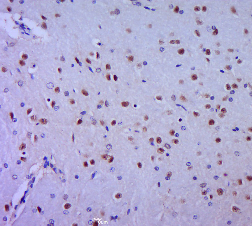
 on human liver. Hes1 was detected in paraffin-embedded section. Heat mediated antigen retrieval was performed in citrate buffer (pH6, epitope retrieval solution) for 20 mins. The tissue section was blocked with 10% goat serum. The tissue section was then incubated with 1ug/ml rabbit anti-Hes1 Antibody (M01459) overnight at 4°C. Biotinylated goat anti Rabbit IgG IgG antibody was used as secondary antibody and incubated for 30 minutes at 37°C. The tissue section was developed using Strepavidin-Biotin-Complex (SABC)(Catalog # SA1022) with DAB as the chromogen.")

Figure 1. Western blot analysis of Hes1 using anti-Hes1 antibody (M01459). Electrophoresis was performed on a 5-20% SDS-PAGE gel at 70V (Stacking gel) / 90V (Resolving gel) for 2-3 hours. The sample well of each lane was loaded with 30 ug of sample under reducing conditions. Lane 1: human U-87MG whole cell lysates, Lane 2: human U251 whole cell lysates, Lane 3: human 293T whole cell lysates, Lane 4: rat brain tissue lysates, Lane 5: mouse brain tissue lysates. After electrophoresis, proteins were transferred to a nitrocellulose membrane at 150 mA for 50-90 minutes. Blocked the membrane with 5% non-fat milk/TBS for 1.5 hour at RT. The membrane was incubated with rabbit anti-Hes1 antigen affinity purified monoclonal antibody (Catalog # M01459) at 1:500 overnight at 4°C, then washed with TBS-0.1%Tween 3 times with 5 minutes each and probed with a goat anti-rabbit IgG-HRP secondary antibody at a dilution of 1:500 for 1.5 hour at RT. The signal is developed using an Enhanced Chemiluminescent detection (ECL) kit (Catalog # EK1002) with Tanon 5200 system. A specific band was detected for Hes1 at approximately 30 kDa. The expected band size for Hes1 is at 30 kDa.
Anti-Hes1 Rabbit Monoclonal Antibody
M01459
ApplicationsFlow Cytometry, ImmunoFluorescence, Western Blot, ImmunoCytoChemistry, ImmunoHistoChemistry
Product group Antibodies
ReactivityHuman, Mouse, Rat
TargetHES1
Overview
- SupplierBoster Bio
- Product NameAnti-Hes1 Rabbit Monoclonal Antibody
- Delivery Days Customer9
- ApplicationsFlow Cytometry, ImmunoFluorescence, Western Blot, ImmunoCytoChemistry, ImmunoHistoChemistry
- CertificationResearch Use Only
- ClonalityMonoclonal
- Clone IDFBB-8
- Gene ID3280
- Target nameHES1
- Target descriptionhes family bHLH transcription factor 1
- Target synonymsHES-1, HHL, HRY, bHLHb39, transcription factor HES-1, class B basic helix-loop-helix protein 39, hairy homolog, hairy-like protein
- HostRabbit
- IsotypeIgG
- Protein IDQ14469
- Protein NameTranscription factor HES-1
- Scientific DescriptionBoster Bio Anti-Hes1 Rabbit Monoclonal Antibody catalog # M01459. Tested in WB, IHC, ICC/IF, Flow Cytometry applications. This antibody reacts with Human, Mouse, Rat.
- ReactivityHuman, Mouse, Rat
- Storage Instruction-20°C
- UNSPSC12352203
References
- Su J, Tao Y, Liu J, et al. Tibetan medicine Qi-Sai-Er-Sang-Dang-Song Decoction inhibits TNF-α-induced rheumatoid arthritis in human fibroblast-like synoviocytes via regulating NOTCH1/NF-κB/NLRP3 pathway. J Ethnopharmacol. 2023,310:116402. doi: 10.1016/j.jep.2023.116402Read this paper
- Shi J, Han G, Wang J, et al. Matrine promotes hepatic oval cells differentiation into hepatocytes and alleviates liver injury by suppression of Notch signalling pathway. Life Sci. 2020,261:118354. doi: 10.1016/j.lfs.2020.118354Read this paper
- Shi J, Han X, Wang J, et al. Matrine prevents the early development of hepatocellular carcinoma like lesions in rat liver. Exp Ther Med. 2019,18(4):2583-2591. doi: 10.3892/etm.2019.7875Read this paper
- Lu L, Liu S, Dong Q, et al. Salidroside suppresses the metastasis of hepatocellular carcinoma cells by inhibiting the activation of the Notch1 signaling pathway. Mol Med Rep. 2019,19(6):4964-4972. doi: 10.3892/mmr.2019.10115Read this paper
- Guan J, Wei X, Qu S, et al. Osthole prevents cerebral ischemia-reperfusion injury via the Notch signaling pathway. Biochem Cell Biol. 2017,95(4):459-467. doi: 10.1139/bcb-2016-0233Read this paper
Datasheet
MSDS
Related products
Product group Antibodies
HES1 AntibodyCSB-PA245265
ApplicationsWestern Blot, ELISA
ReactivityHuman, Mouse, Rat
TargetHES1
- SizePrice
Product group Antibodies

HES1 / HES-1 AntibodyLS-C747087
ApplicationsWestern Blot
ReactivityHuman, Rat
TargetHES1
- SizePrice

Product group Antibodies
Anti-HES1 AntibodyHPA066929
ApplicationsWestern Blot, ImmunoCytoChemistry
ReactivityHuman
TargetHES1
- SizePrice
Product group Antibodies
ApplicationsImmunoPrecipitation, Western Blot, ImmunoCytoChemistry, ImmunoHistoChemistry
ReactivityMouse
TargetHES1
- SizePrice
Product group Antibodies
References
HES1 Polyclonal AntibodyBS-2972R
ApplicationsFlow Cytometry, ImmunoFluorescence, Western Blot, ELISA, ImmunoCytoChemistry, ImmunoHistoChemistry, ImmunoHistoChemistry Frozen, ImmunoHistoChemistry Paraffin
ReactivityBovine, Equine, Human, Mouse, Porcine, Rabbit, Rat
TargetHES1
- SizePrice
![Various whole cell extracts (30 μg) were separated by 12% SDS-PAGE, and the membrane was blotted with HES1 antibody [N1], N-term (GTX108356) diluted at 1:1000. The HRP-conjugated anti-rabbit IgG antibody (GTX213110-01) was used to detect the primary antibody. Corresponding RNA expression data for the same cell lines are based on Human Protein Atlas program.](https://www.genetex.com/upload/website/prouct_img/normal/GTX108356/GTX108356_44860_20221118_WB_TPM_watermark_23030219_766.webp)
Product group Antibodies
HES1 antibody [N1], N-termGTX108356
ApplicationsImmunoFluorescence, ImmunoPrecipitation, Western Blot, ImmunoCytoChemistry
ReactivityHuman, Mouse, Rat
TargetHES1
- SizePrice
Product group Antibodies
Anti-HES1 Antibody144-62113
ApplicationsWestern Blot
ReactivityHuman, Rat
TargetHES1
- SizePrice