


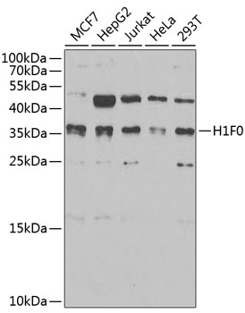
Figure 1. Western blot analysis of Histone H1.0/H1F0 using anti-Histone H1.0/H1F0 antibody (M08821-2). Electrophoresis was performed on a 5-20% SDS-PAGE gel at 70V (Stacking gel) / 90V (Resolving gel) for 2-3 hours. The sample well of each lane was loaded with 30 ug of sample under reducing conditions. Lane 1: human U20S whole cell lysates, Lane 2: human PC-3 whole cell lysates, Lane 3: human HepG2 whole cell lysates. After electrophoresis, proteins were transferred to a nitrocellulose membrane at 150 mA for 50-90 minutes. Blocked the membrane with 5% non-fat milk/TBS for 1.5 hour at RT. The membrane was incubated with mouse anti-Histone H1.0/H1F0 antigen affinity purified monoclonal antibody (Catalog # M08821-2) at 0.5 microg/mL overnight at 4°C, then washed with TBS-0.1%Tween 3 times with 5 minutes each and probed with a goat anti-mouse IgG-HRP secondary antibody at a dilution of 1:10000 for 1.5 hour at RT. The signal is developed using an Enhanced Chemiluminescent detection (ECL) kit (Catalog # EK1001) with Tanon 5200 system. A specific band was detected for Histone H1.0/H1F0 at approximately 24 kDa. The expected band size for Histone H1.0/H1F0 is at 24 kDa.
. Histone H1.0/H1F0 was detected in a paraffin-embedded section of mouse colon tissue. Heat mediated antigen retrieval was performed in EDTA buffer (pH 8.0, epitope retrieval solution). The tissue section was blocked with 10% goat serum. The tissue section was then incubated with 2 microg/ml mouse anti-Histone H1.0/H1F0 Antibody (M08821-2) overnight at 4°C. Peroxidase Conjugated Goat Anti-mouse IgG was used as secondary antibody and incubated for 30 minutes at 37°C. The tissue section was developed using HRP Conjugated Mouse IgG Super Vision Assay Kit (Catalog # SV0001) with DAB as the chromogen.")
. Histone H1.0/H1F0 was detected in a paraffin-embedded section of human placenta tissue. Heat mediated antigen retrieval was performed in EDTA buffer (pH 8.0, epitope retrieval solution). The tissue section was blocked with 10% goat serum. The tissue section was then incubated with 2 microg/ml mouse anti-Histone H1.0/H1F0 Antibody (M08821-2) overnight at 4°C. Peroxidase Conjugated Goat Anti-mouse IgG was used as secondary antibody and incubated for 30 minutes at 37°C. The tissue section was developed using HRP Conjugated Mouse IgG Super Vision Assay Kit (Catalog # SV0001) with DAB as the chromogen.")
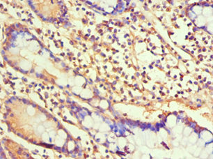
. Histone H1.0/H1F0 was detected in a paraffin-embedded section of human differentiated adenocarcinoma of the rectum tissue. Heat mediated antigen retrieval was performed in EDTA buffer (pH 8.0, epitope retrieval solution). The tissue section was blocked with 10% goat serum. The tissue section was then incubated with 2 microg/ml mouse anti-Histone H1.0/H1F0 Antibody (M08821-2) overnight at 4°C. Peroxidase Conjugated Goat Anti-mouse IgG was used as secondary antibody and incubated for 30 minutes at 37°C. The tissue section was developed using HRP Conjugated Mouse IgG Super Vision Assay Kit (Catalog # SV0001) with DAB as the chromogen.")
. Histone H1.0/H1F0 was detected in a paraffin-embedded section of human Hodgkins lymphoma tissue. Heat mediated antigen retrieval was performed in EDTA buffer (pH 8.0, epitope retrieval solution). The tissue section was blocked with 10% goat serum. The tissue section was then incubated with 2 microg/ml mouse anti-Histone H1.0/H1F0 Antibody (M08821-2) overnight at 4°C. Peroxidase Conjugated Goat Anti-mouse IgG was used as secondary antibody and incubated for 30 minutes at 37°C. The tissue section was developed using HRP Conjugated Mouse IgG Super Vision Assay Kit (Catalog # SV0001) with DAB as the chromogen.")
. Histone H1.0/H1F0 was detected in a paraffin-embedded section of human renal cell carcinoma tissue. Heat mediated antigen retrieval was performed in EDTA buffer (pH 8.0, epitope retrieval solution). The tissue section was blocked with 10% goat serum. The tissue section was then incubated with 2 microg/ml mouse anti-Histone H1.0/H1F0 Antibody (M08821-2) overnight at 4°C. Peroxidase Conjugated Goat Anti-mouse IgG was used as secondary antibody and incubated for 30 minutes at 37°C. The tissue section was developed using HRP Conjugated Mouse IgG Super Vision Assay Kit (Catalog # SV0001) with DAB as the chromogen.")
. Histone H1.0/H1F0 was detected in a paraffin-embedded section of human tonsil tissue. Heat mediated antigen retrieval was performed in EDTA buffer (pH 8.0, epitope retrieval solution). The tissue section was blocked with 10% goat serum. The tissue section was then incubated with 2 microg/ml mouse anti-Histone H1.0/H1F0 Antibody (M08821-2) overnight at 4°C. Peroxidase Conjugated Goat Anti-mouse IgG was used as secondary antibody and incubated for 30 minutes at 37°C. The tissue section was developed using HRP Conjugated Mouse IgG Super Vision Assay Kit (Catalog # SV0001) with DAB as the chromogen.")
. Histone H1.0/H1F0 was detected in a paraffin-embedded section of human gastric carcinoma tissue. Heat mediated antigen retrieval was performed in EDTA buffer (pH 8.0, epitope retrieval solution). The tissue section was blocked with 10% goat serum. The tissue section was then incubated with 2 microg/ml mouse anti-Histone H1.0/H1F0 Antibody (M08821-2) overnight at 4°C. Peroxidase Conjugated Goat Anti-mouse IgG was used as secondary antibody and incubated for 30 minutes at 37°C. The tissue section was developed using HRP Conjugated Mouse IgG Super Vision Assay Kit (Catalog # SV0001) with DAB as the chromogen.")
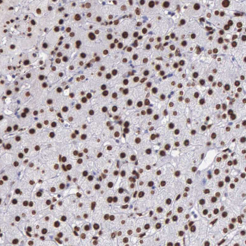
. Histone H1.0/H1F0 was detected in a paraffin-embedded section of human liver cancer tissue. Heat mediated antigen retrieval was performed in EDTA buffer (pH 8.0, epitope retrieval solution). The tissue section was blocked with 10% goat serum. The tissue section was then incubated with 2 microg/ml mouse anti-Histone H1.0/H1F0 Antibody (M08821-2) overnight at 4°C. Peroxidase Conjugated Goat Anti-mouse IgG was used as secondary antibody and incubated for 30 minutes at 37°C. The tissue section was developed using HRP Conjugated Mouse IgG Super Vision Assay Kit (Catalog # SV0001) with DAB as the chromogen.")
. Overlay histogram showing SiHa cells stained with M08821-2 (Blue line). To facilitate intracellular staining, cells were fixed with 4% paraformaldehyde and permeabilized with permeabilization buffer. The cells were blocked with 10% normal goat serum. And then incubated with mouse anti-Histone H1.0/H1F0 Antibody (M08821-2, 1 microg/1x106 cells) for 30 min at 20°C. DyLight®488 conjugated goat anti-mouse IgG (BA1126, 5-10 microg/1x106 cells) was used as secondary antibody for 30 minutes at 20°C. Isotype control antibody (Green line) was mouse IgG (1 microg/1x106) used under the same conditions. Unlabelled sample without incubation with primary antibody and secondary antibody (Red line) was used as a blank control.")
Figure 1. Western blot analysis of Histone H1.0/H1F0 using anti-Histone H1.0/H1F0 antibody (M08821-2). Electrophoresis was performed on a 5-20% SDS-PAGE gel at 70V (Stacking gel) / 90V (Resolving gel) for 2-3 hours. The sample well of each lane was loaded with 30 ug of sample under reducing conditions. Lane 1: human U20S whole cell lysates, Lane 2: human PC-3 whole cell lysates, Lane 3: human HepG2 whole cell lysates. After electrophoresis, proteins were transferred to a nitrocellulose membrane at 150 mA for 50-90 minutes. Blocked the membrane with 5% non-fat milk/TBS for 1.5 hour at RT. The membrane was incubated with mouse anti-Histone H1.0/H1F0 antigen affinity purified monoclonal antibody (Catalog # M08821-2) at 0.5 microg/mL overnight at 4°C, then washed with TBS-0.1%Tween 3 times with 5 minutes each and probed with a goat anti-mouse IgG-HRP secondary antibody at a dilution of 1:10000 for 1.5 hour at RT. The signal is developed using an Enhanced Chemiluminescent detection (ECL) kit (Catalog # EK1001) with Tanon 5200 system. A specific band was detected for Histone H1.0/H1F0 at approximately 24 kDa. The expected band size for Histone H1.0/H1F0 is at 24 kDa.
Anti-Histone H1.0/H1F0 Antibody Picoband(r) (monoclonal, 5I3E6)
M08821-2-IFLUOR647
ApplicationsFlow Cytometry, Western Blot, ImmunoHistoChemistry
Product group Antibodies
ReactivityHuman, Mouse
TargetH1-0
Overview
- SupplierBoster Bio
- Product NameAnti-Histone H1.0/H1F0 Antibody Picoband(r) (monoclonal, 5I3E6)
- Delivery Days Customer9
- ApplicationsFlow Cytometry, Western Blot, ImmunoHistoChemistry
- CertificationResearch Use Only
- ClonalityMonoclonal
- Clone ID5I3E6
- Concentration500 ug/ml
- ConjugateOther Conjugate
- Gene ID3005
- Target nameH1-0
- Target descriptionH1.0 linker histone
- Target synonymsH1.0, H10, H1F0, H1FV, histone H1.0, H1 histone family member 0, H1.0, H1(0), H1-0, histone H1', histone H1(0)
- HostMouse
- IsotypeIgG2b
- Protein IDP07305
- Protein NameHistone H1.0
- Scientific DescriptionBoster Bio Anti-Histone H1.0/H1F0 Antibody Picoband® (monoclonal, 5I3E6) catalog # M08821-2. Tested in Flow Cytometry, IHC, WB applications. This antibody reacts with Human, Mouse. The brand Picoband indicates this is a premium antibody that guarantees superior quality, high affinity, and strong signals with minimal background in Western blot applications. Only our best-performing antibodies are designated as Picoband, ensuring unmatched performance.
- ReactivityHuman, Mouse
- Storage Instruction-20°C,2°C to 8°C
- UNSPSC12352203
Related products

Product group Antibodies
Anti-H1F0 Antibody144-03298
ApplicationsImmunoFluorescence, Western Blot, ImmunoHistoChemistry
ReactivityHuman, Mouse, Rat
TargetH1-0
- SizePrice
Product group Antibodies
H1F0 (Ab-11) Polyclonal AntibodyCAC15243
ApplicationsWestern Blot, ELISA
ReactivityRat
TargetH1-0
- SizePrice
Product group Antibodies
Histone H1.0 Polyclonal AntibodyBS-33535R
ApplicationsWestern Blot, ELISA
ReactivityHuman, Mouse, Rat
TargetH1-0
- SizePrice
Product group Antibodies
H1F0 AntibodyCSB-PA010087ESR1HU
ApplicationsELISA, ImmunoHistoChemistry
ReactivityHuman
TargetH1-0
- SizePrice
Product group Antibodies
ReactivityHuman
TargetH1-0
- SizePrice
Product group Antibodies
ApplicationsImmunoFluorescence, Western Blot, ImmunoCytoChemistry, ImmunoHistoChemistry
ReactivityHuman, Mouse, Rat
- SizePrice
Product group Antibodies
Anti-H1F0 AntibodyHPA000843
ApplicationsWestern Blot, ImmunoCytoChemistry, ImmunoHistoChemistry
ReactivityHuman
TargetH1-0
- SizePrice
Product group Antibodies

H1F0 AntibodyLS-C409090
ApplicationsWestern Blot
ReactivityHuman
TargetH1-0
- SizePrice