

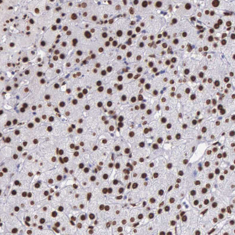
Immunohistochemical staining of human liver shows strong nuclear positivity in hepatocytes.
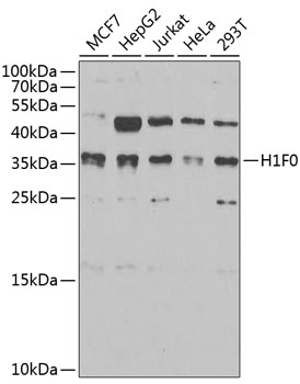
![Lane 1: Marker [kDa] 250, 130, 100, 70, 55, 35, 25, 15, 10. Lane 2: Human cell line U-2 OS](https://atlasantibodies.s3.amazonaws.com/images/wb/hpa000843-wb-1.jpg "Lane 1: Marker [kDa] 250, 130, 100, 70, 55, 35, 25, 15, 10. Lane 2: Human cell line U-2 OS")
Immunohistochemical staining of human liver shows strong nuclear positivity in hepatocytes.
Anti-H1F0 Antibody
HPA000843
ApplicationsWestern Blot, ImmunoCytoChemistry, ImmunoHistoChemistry
Product group Antibodies
ReactivityHuman
TargetH1-0
Overview
- SupplierAtlas Antibodies
- Product NameAnti-H1F0 Antibody
- Delivery Days Customer4
- ApplicationsWestern Blot, ImmunoCytoChemistry, ImmunoHistoChemistry
- CertificationResearch Use Only
- ClonalityPolyclonal
- ConjugateUnconjugated
- Gene ID3005
- Target nameH1-0
- Target descriptionH1.0 linker histone
- Target synonymsH1.0, H10, H1F0, H1FV, histone H1.0, H1 histone family member 0, H1.0, H1(0), H1-0, histone H1', histone H1(0)
- HostRabbit
- IsotypeIgG
- Protein IDP07305
- Protein NameHistone H1.0
- Scientific DescriptionRecombinant Protein Epitope Signature Tag (PrEST) antigen sequence
- ReactivityHuman
- Storage Instruction-20°C,2°C to 8°C
- UNSPSC41116161
Datasheet
MSDS
Related products
Product group Antibodies
ApplicationsImmunoFluorescence, Western Blot, ImmunoCytoChemistry, ImmunoHistoChemistry
ReactivityHuman, Mouse, Rat
- SizePrice


Product group Antibodies
Anti-Histone H1.0/H1F0 Antibody Picoband(r)A08821-1-CARRIER-FREE
ApplicationsFlow Cytometry, ImmunoFluorescence, Western Blot, ELISA, ImmunoCytoChemistry, ImmunoHistoChemistry
ReactivityHuman, Mouse, Rat
TargetH1-0
- SizePrice
Product group Antibodies
Anti-H1F0 Antibody144-03298
ApplicationsImmunoFluorescence, Western Blot, ImmunoHistoChemistry
ReactivityHuman, Mouse, Rat
TargetH1-0
- SizePrice
Product group Antibodies
Histone H1.0 Polyclonal AntibodyBS-33535R
ApplicationsWestern Blot, ELISA
ReactivityHuman, Mouse, Rat
TargetH1-0
- SizePrice
Product group Antibodies
H1F0 AntibodyCSB-PA010087ESR1HU
ApplicationsELISA, ImmunoHistoChemistry
ReactivityHuman
TargetH1-0
- SizePrice
Product group Antibodies
H1F0 (Ab-11) Polyclonal AntibodyCAC15243
ApplicationsWestern Blot, ELISA
ReactivityRat
TargetH1-0
- SizePrice
Product group Antibodies

H1F0 AntibodyLS-C409090
ApplicationsWestern Blot
ReactivityHuman
TargetH1-0
- SizePrice
Product group Antibodies
Histone H1.0 antibodyGTX114462
ApplicationsImmunoFluorescence, Western Blot, ImmunoCytoChemistry, ImmunoHistoChemistry, ImmunoHistoChemistry Paraffin
ReactivityHuman, Mouse, Rat
TargetH1-0
- SizePrice