

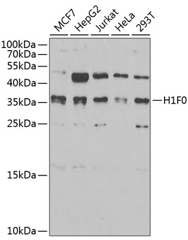

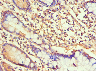
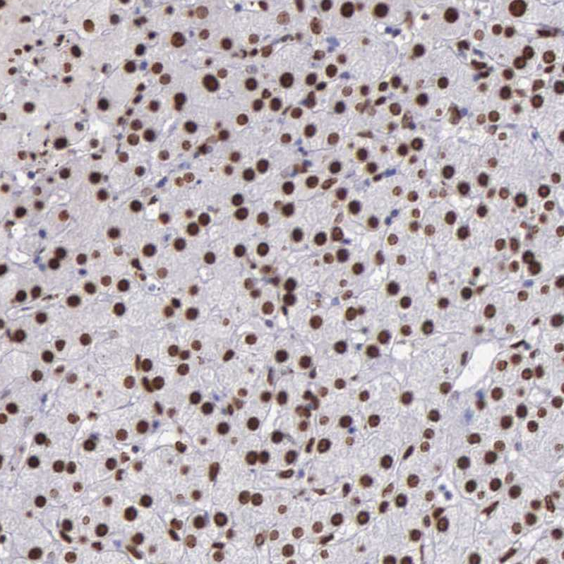
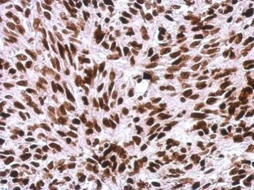
Anti-Histone H1.0 Antibody
A15429
ApplicationsImmunoFluorescence, Western Blot, ImmunoCytoChemistry, ImmunoHistoChemistry
Product group Antibodies
ReactivityHuman, Mouse, Rat
Overview
- SupplierAntibodies.com
- Product NameAnti-Histone H1.0 Antibody
- Delivery Days Customer7
- ApplicationsImmunoFluorescence, Western Blot, ImmunoCytoChemistry, ImmunoHistoChemistry
- CertificationResearch Use Only
- ClonalityPolyclonal
- ConjugateUnconjugated
- HostRabbit
- IsotypeIgG
- Scientific DescriptionRabbit polyclonal antibody to Histone H1.0.
- ReactivityHuman, Mouse, Rat
- UNSPSC12352203
Related products


Product group Antibodies
Anti-Histone H1.0/H1F0 Antibody Picoband(r)A08821-1-CARRIER-FREE
ApplicationsFlow Cytometry, ImmunoFluorescence, Western Blot, ELISA, ImmunoCytoChemistry, ImmunoHistoChemistry
ReactivityHuman, Mouse, Rat
TargetH1-0
- SizePrice
Product group Antibodies
Anti-H1F0 Antibody144-03298
ApplicationsImmunoFluorescence, Western Blot, ImmunoHistoChemistry
ReactivityHuman, Mouse, Rat
TargetH1-0
- SizePrice
Product group Antibodies
Histone H1.0 Polyclonal AntibodyBS-33535R
ApplicationsWestern Blot, ELISA
ReactivityHuman, Mouse, Rat
TargetH1-0
- SizePrice
Product group Antibodies
H1F0 AntibodyCSB-PA010087ESR1HU
ApplicationsELISA, ImmunoHistoChemistry
ReactivityHuman
TargetH1-0
- SizePrice
Product group Antibodies
H1F0 (Ab-11) Polyclonal AntibodyCAC15243
ApplicationsWestern Blot, ELISA
ReactivityRat
TargetH1-0
- SizePrice
Product group Antibodies

H1F0 AntibodyLS-C409090
ApplicationsWestern Blot
ReactivityHuman
TargetH1-0
- SizePrice
Product group Antibodies
Anti-H1F0 AntibodyHPA000843
ApplicationsWestern Blot, ImmunoCytoChemistry, ImmunoHistoChemistry
ReactivityHuman
TargetH1-0
- SizePrice
Product group Antibodies
Histone H1.0 antibodyGTX114462
ApplicationsImmunoFluorescence, Western Blot, ImmunoCytoChemistry, ImmunoHistoChemistry, ImmunoHistoChemistry Paraffin
ReactivityHuman, Mouse, Rat
TargetH1-0
- SizePrice
Product group Antibodies
ApplicationsWestern Blot, ELISA, ImmunoHistoChemistry, ImmunoHistoChemistry Paraffin
ReactivityHuman
TargetH1-0
- SizePrice