


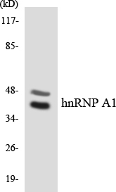
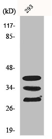
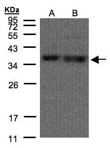
Figure 1. Western blot analysis of HnRNP A1 using anti-HnRNP A1 antibody (A01476). Electrophoresis was performed on a 5-20% SDS-PAGE gel at 70V (Stacking gel) / 90V (Resolving gel) for 2-3 hours. The sample well of each lane was loaded with 30 ug of sample under reducing conditions. Lane 1: human HepG2 whole cell lysates, Lane 2: human Daudi whole cell lysates, Lane 3: human MOLT-4 whole cell lysates, Lane 4: human HL-60 whole cell lysates. After electrophoresis, proteins were transferred to a nitrocellulose membrane at 150 mA for 50-90 minutes. Blocked the membrane with 5% non-fat milk/TBS for 1.5 hour at RT. The membrane was incubated with rabbit anti-HnRNP A1 antigen affinity purified polyclonal antibody (Catalog # A01476) at 0.5 microg/mL overnight at 4°C, then washed with TBS-0.1%Tween 3 times with 5 minutes each and probed with a goat anti-rabbit IgG-HRP secondary antibody at a dilution of 1:5000 for 1.5 hour at RT. The signal is developed using an Enhanced Chemiluminescent detection (ECL) kit (Catalog # EK1002) with Tanon 5200 system. A specific band was detected for HnRNP A1 at approximately 36 kDa. The expected band size for HnRNP A1 is at 39 kDa.
. HnRNP A1 was detected in paraffin-embedded section of mouse intestine tissues. Heat mediated antigen retrieval was performed in citrate buffer (pH6, epitope retrieval solution) for 20 mins. The tissue section was blocked with 10% goat serum. The tissue section was then incubated with 1microg/ml rabbit anti-HnRNP A1 Antibody (A01476) overnight at 4°C. Biotinylated goat anti-rabbit IgG was used as secondary antibody and incubated for 30 minutes at 37°C. The tissue section was developed using Strepavidin-Biotin-Complex (SABC)(Catalog # SA1022) with DAB as the chromogen.")
. HnRNP A1 was detected in paraffin-embedded section of mouse kidney tissues. Heat mediated antigen retrieval was performed in citrate buffer (pH6, epitope retrieval solution) for 20 mins. The tissue section was blocked with 10% goat serum. The tissue section was then incubated with 1microg/ml rabbit anti-HnRNP A1 Antibody (A01476) overnight at 4°C. Biotinylated goat anti-rabbit IgG was used as secondary antibody and incubated for 30 minutes at 37°C. The tissue section was developed using Strepavidin-Biotin-Complex (SABC)(Catalog # SA1022) with DAB as the chromogen.")
. HnRNP A1 was detected in paraffin-embedded section of rat kidney tissues. Heat mediated antigen retrieval was performed in citrate buffer (pH6, epitope retrieval solution) for 20 mins. The tissue section was blocked with 10% goat serum. The tissue section was then incubated with 1microg/ml rabbit anti-HnRNP A1 Antibody (A01476) overnight at 4°C. Biotinylated goat anti-rabbit IgG was used as secondary antibody and incubated for 30 minutes at 37°C. The tissue section was developed using Strepavidin-Biotin-Complex (SABC)(Catalog # SA1022) with DAB as the chromogen.")
. HnRNP A1 was detected in paraffin-embedded section of human intestinal cancer tissues. Heat mediated antigen retrieval was performed in citrate buffer (pH6, epitope retrieval solution) for 20 mins. The tissue section was blocked with 10% goat serum. The tissue section was then incubated with 1microg/ml rabbit anti-HnRNP A1 Antibody (A01476) overnight at 4°C. Biotinylated goat anti-rabbit IgG was used as secondary antibody and incubated for 30 minutes at 37°C. The tissue section was developed using Strepavidin-Biotin-Complex (SABC)(Catalog # SA1022) with DAB as the chromogen.")
. HnRNP A1 was detected in paraffin-embedded section of human placenta tissues. Heat mediated antigen retrieval was performed in citrate buffer (pH6, epitope retrieval solution) for 20 mins. The tissue section was blocked with 10% goat serum. The tissue section was then incubated with 1microg/ml rabbit anti-HnRNP A1 Antibody (A01476) overnight at 4°C. Biotinylated goat anti-rabbit IgG was used as secondary antibody and incubated for 30 minutes at 37°C. The tissue section was developed using Strepavidin-Biotin-Complex (SABC)(Catalog # SA1022) with DAB as the chromogen.")
. HnRNP A1 was detected in paraffin-embedded section of human rectal cancer tissue. Heat mediated antigen retrieval was performed in EDTA buffer (pH8.0, epitope retrieval solution). The tissue section was blocked with 10% goat serum. The tissue section was then incubated with 5microg/mL rabbit anti-HnRNP A1 Antibody (A01476) overnight at 4°C. DyLight®488 Conjugated Goat Anti-Rabbit IgG (BA1127) was used as secondary antibody at 1:100 dilution and incubated for 30 minutes at 37°C. The section was counterstained with DAPI. Visualize using a fluorescence microscope and filter sets appropriate for the label used.")
. HnRNP A1 was detected in a paraffin-embedded section of mouse brain tissue. Heat mediated antigen retrieval was performed in EDTA buffer (pH 8.0, epitope retrieval solution). The tissue section was blocked with 10% goat serum. The tissue section was then incubated with 5 microg/mL rabbit anti-HnRNP A1 Antibody (A01476) overnight at 4°C. DyLight488 Conjugated Goat Anti-Rabbit IgG (BA1127) was used as secondary antibody at 1:500 dilution and incubated for 30 minutes at 37°C. Visualize using a fluorescence microscope and filter sets appropriate for the label used.")
. HnRNP A1 was detected in a paraffin-embedded section of rat brain tissue. Heat mediated antigen retrieval was performed in EDTA buffer (pH 8.0, epitope retrieval solution). The tissue section was blocked with 10% goat serum. The tissue section was then incubated with 5 microg/mL rabbit anti-HnRNP A1 Antibody (A01476) overnight at 4°C. DyLight488 Conjugated Goat Anti-Rabbit IgG (BA1127) was used as secondary antibody at 1:500 dilution and incubated for 30 minutes at 37°C. Visualize using a fluorescence microscope and filter sets appropriate for the label used.")
. HnRNP A1 was detected in immunocytochemical section of U20S cells. Enzyme antigen retrieval was performed using IHC enzyme antigen retrieval reagent (AR0022) for 15 mins. The cells were blocked with 10% goat serum. And then incubated with 2microg/mL rabbit anti-HnRNP A1 Antibody (A01476) overnight at 4°C. DyLight®550 Conjugated Goat Anti-Rabbit IgG (BA1135) was used as secondary antibody at 1:100 dilution and incubated for 30 minutes at 37°C. The section was counterstained with DAPI. Visualize using a fluorescence microscope and filter sets appropriate for the label used.")
Figure 1. Western blot analysis of HnRNP A1 using anti-HnRNP A1 antibody (A01476). Electrophoresis was performed on a 5-20% SDS-PAGE gel at 70V (Stacking gel) / 90V (Resolving gel) for 2-3 hours. The sample well of each lane was loaded with 30 ug of sample under reducing conditions. Lane 1: human HepG2 whole cell lysates, Lane 2: human Daudi whole cell lysates, Lane 3: human MOLT-4 whole cell lysates, Lane 4: human HL-60 whole cell lysates. After electrophoresis, proteins were transferred to a nitrocellulose membrane at 150 mA for 50-90 minutes. Blocked the membrane with 5% non-fat milk/TBS for 1.5 hour at RT. The membrane was incubated with rabbit anti-HnRNP A1 antigen affinity purified polyclonal antibody (Catalog # A01476) at 0.5 microg/mL overnight at 4°C, then washed with TBS-0.1%Tween 3 times with 5 minutes each and probed with a goat anti-rabbit IgG-HRP secondary antibody at a dilution of 1:5000 for 1.5 hour at RT. The signal is developed using an Enhanced Chemiluminescent detection (ECL) kit (Catalog # EK1002) with Tanon 5200 system. A specific band was detected for HnRNP A1 at approximately 36 kDa. The expected band size for HnRNP A1 is at 39 kDa.
Anti-HnRNP A1/HNRNPA1 Antibody Picoband(r)
A01476-CARRIER-FREE
ApplicationsFlow Cytometry, ImmunoFluorescence, Western Blot, ImmunoCytoChemistry, ImmunoHistoChemistry
Product group Antibodies
ReactivityHuman, Mouse, Rat
TargetHNRNPA1
Overview
- SupplierBoster Bio
- Product NameAnti-HnRNP A1/HNRNPA1 Antibody Picoband(r)
- Delivery Days Customer9
- Application Supplier NoteTested Species: In-house tested species with positive results. By Heat: Boiling the paraffin sections in 10mM citrate buffer, pH6.0, for 20mins is required for the staining of formalin/paraffin sections. Other applications have not been tested. Optimal dilutions should be determined by end users.
- ApplicationsFlow Cytometry, ImmunoFluorescence, Western Blot, ImmunoCytoChemistry, ImmunoHistoChemistry
- CertificationResearch Use Only
- ClonalityPolyclonal
- Concentration500 ug/ml
- Gene ID3178
- Target nameHNRNPA1
- Target descriptionheterogeneous nuclear ribonucleoprotein A1
- Target synonymsALS19, ALS20, HNRPA1, HNRPA1L3, IBMPFD3, MPD3, UP 1, hnRNP A1, hnRNP-A1, heterogeneous nuclear ribonucleoprotein A1, epididymis secretory sperm binding protein, helix-destabilizing protein, heterogeneous nuclear ribonucleoprotein A1B protein, heterogeneous nuclear ribonucleoprotein B2 protein, heterogeneous nuclear ribonucleoprotein core protein A1, hnRNP core protein A1-like 3, nuclear ribonucleoprotein particle A1 protein, putative heterogeneous nuclear ribonucleoprotein A1-like 3, single-strand DNA-binding protein UP1, single-strand RNA-binding protein
- HostRabbit
- IsotypeIgG
- Protein IDP09651
- Protein NameHeterogeneous nuclear ribonucleoprotein A1
- Scientific DescriptionBoster Bio Anti-HnRNP A1/HNRNPA1 Antibody Picoband® catalog # A01476. Tested in Flow Cytometry, IF, IHC, ICC, WB applications. This antibody reacts with Human, Mouse, Rat. The brand Picoband indicates this is a premium antibody that guarantees superior quality, high affinity, and strong signals with minimal background in Western blot applications. Only our best-performing antibodies are designated as Picoband, ensuring unmatched performance.
- ReactivityHuman, Mouse, Rat
- Storage Instruction-20°C,2°C to 8°C
- UNSPSC12352203
Related products
Product group Antibodies
Anti-hnRNP A1 AntibodyA95344
ApplicationsWestern Blot, ELISA, ImmunoHistoChemistry
ReactivityHuman, Mouse, Rat
- SizePrice
Product group Antibodies
Anti-HNRNPA1 Antibody144-07491
ApplicationsImmunoFluorescence, Western Blot, ImmunoHistoChemistry
ReactivityHuman, Mouse, Rat
TargetHNRNPA1
- SizePrice
Product group Antibodies
hnRNP A1 Recombinant Antibody, AbBy Fluor-488 ConjugatedBSM-61490R-BF488
ApplicationsFlow Cytometry, ImmunoFluorescence, Western Blot
ReactivityHuman, Mouse, Rat
TargetHNRNPA1
- SizePrice
Product group Antibodies
HNRNPA1 AntibodyCSB-PA002942
ApplicationsImmunoFluorescence, Western Blot, ELISA, ImmunoHistoChemistry
ReactivityHuman, Mouse, Rat
TargetHNRNPA1
- SizePrice
Product group Antibodies
Hnrnpa1 Polyclonal AntibodyCAC07236
ApplicationsImmunoFluorescence, Western Blot, ELISA, ImmunoHistoChemistry
TargetHNRNPA1
- SizePrice

Product group Antibodies
Anti-HNRNPA1 AntibodyHPA001609
ApplicationsWestern Blot, ImmunoHistoChemistry
ReactivityHuman
TargetHNRNPA1
- SizePrice
Product group Antibodies
hnRNP A1 antibodyGTX106208
ApplicationsImmunoFluorescence, Western Blot, ImmunoCytoChemistry, ImmunoHistoChemistry, ImmunoHistoChemistry Paraffin
ReactivityHuman, Mouse, Rat
TargetHNRNPA1
- SizePrice
Product group Antibodies

HNRPA1 / HnRNP A1 AntibodyLS-C747546
ApplicationsImmunoFluorescence, Western Blot
ReactivityHuman, Mouse, Rat
TargetHNRNPA1
- SizePrice