


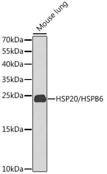
Figure 1. Western blot analysis of Hsp20 using anti-Hsp20 antibody (A07981-1). Electrophoresis was performed on a 5-20% SDS-PAGE gel at 70V (Stacking gel) / 90V (Resolving gel) for 2-3 hours. The sample well of each lane was loaded with 50ug of sample under reducing conditions. Lane 1: rat cardiac muscle tissue lysate,Lane 2: rat skeletal muscle tissue lysate,Lane 3: rat testis tissue lysate,Lane 4: mouse testis tissue lysate,Lane 5: human A431 whole Cell lysate.After Electrophoresis, proteins were transferred to a Nitrocellulose membrane at 150mA for 50-90 minutes. Blocked the membrane with 5% Non-fat Milk/ TBS for 1.5 hour at RT. The membrane was incubated with rabbit anti-Hsp20 antigen affinity purified polyclonal antibody (Catalog # A07981-1) at 0.5 microg/mL overnight at 4°C, then washed with TBS-0.1%Tween 3 times with 5 minutes each and probed with a goat anti-rabbit IgG-HRP secondary antibody at a dilution of 1:10000 for 1.5 hour at RT. The signal is developed using an Enhanced Chemiluminescent detection (ECL) kit (Catalog # EK1002) with Tanon 5200 system. A specific band was detected for Hsp20 at approximately 17KD. The expected band size for Hsp20 is at 17KD.
. Hsp20 was detected in paraffin-embedded section of human skeletal muscle tissue. Heat mediated antigen retrieval was performed in EDTA buffer (pH8.0, epitope retrieval solution). The tissue section was blocked with 10% goat serum. The tissue section was then incubated with 1microg/ml rabbit anti-Hsp20 Antibody (A07981-1) overnight at 4°C. Biotinylated goat anti-rabbit IgG was used as secondary antibody and incubated for 30 minutes at 37°C. The tissue section was developed using Strepavidin-Biotin-Complex (SABC) (Catalog # SA1022) with DAB as the chromogen.")
. Hsp20 was detected in paraffin-embedded section of rat cardiac muscle tissue. Heat mediated antigen retrieval was performed in EDTA buffer (pH8.0, epitope retrieval solution). The tissue section was blocked with 10% goat serum. The tissue section was then incubated with 1microg/ml rabbit anti-Hsp20 Antibody (A07981-1) overnight at 4°C. Biotinylated goat anti-rabbit IgG was used as secondary antibody and incubated for 30 minutes at 37°C. The tissue section was developed using Strepavidin-Biotin-Complex (SABC) (Catalog # SA1022) with DAB as the chromogen.")
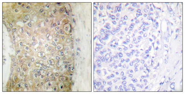
. Hsp20 was detected in paraffin-embedded section of human mammary cancer tissue. Heat mediated antigen retrieval was performed in EDTA buffer (pH8.0, epitope retrieval solution). The tissue section was blocked with 10% goat serum. The tissue section was then incubated with 1microg/ml rabbit anti-Hsp20 Antibody (A07981-1) overnight at 4°C. Biotinylated goat anti-rabbit IgG was used as secondary antibody and incubated for 30 minutes at 37°C. The tissue section was developed using Strepavidin-Biotin-Complex (SABC) (Catalog # SA1022) with DAB as the chromogen.")
. Hsp20 was detected in paraffin-embedded section of mouse lung tissue. Heat mediated antigen retrieval was performed in EDTA buffer (pH8.0, epitope retrieval solution). The tissue section was blocked with 10% goat serum. The tissue section was then incubated with 1microg/ml rabbit anti-Hsp20 Antibody (A07981-1) overnight at 4°C. Biotinylated goat anti-rabbit IgG was used as secondary antibody and incubated for 30 minutes at 37°C. The tissue section was developed using Strepavidin-Biotin-Complex (SABC) (Catalog # SA1022) with DAB as the chromogen.")
. Hsp20 was detected in paraffin-embedded section of mouse cardiac muscle tissue. Heat mediated antigen retrieval was performed in EDTA buffer (pH8.0, epitope retrieval solution). The tissue section was blocked with 10% goat serum. The tissue section was then incubated with 1microg/ml rabbit anti-Hsp20 Antibody (A07981-1) overnight at 4°C. Biotinylated goat anti-rabbit IgG was used as secondary antibody and incubated for 30 minutes at 37°C. The tissue section was developed using Strepavidin-Biotin-Complex (SABC) (Catalog # SA1022) with DAB as the chromogen.")
. Hsp20 was detected in paraffin-embedded section of rat lung tissue. Heat mediated antigen retrieval was performed in EDTA buffer (pH8.0, epitope retrieval solution). The tissue section was blocked with 10% goat serum. The tissue section was then incubated with 1microg/ml rabbit anti-Hsp20 Antibody (A07981-1) overnight at 4°C. Biotinylated goat anti-rabbit IgG was used as secondary antibody and incubated for 30 minutes at 37°C. The tissue section was developed using Strepavidin-Biotin-Complex (SABC) (Catalog # SA1022) with DAB as the chromogen.")
. Hsp20 was detected in immunocytochemical section of A431 cells. Enzyme antigen retrieval was performed using IHC enzyme antigen retrieval reagent (AR0022) for 15 mins. The cells were blocked with 10% goat serum. And then incubated with 2microg/mL rabbit anti-Hsp20 Antibody (A07981-1) overnight at 4°C. DyLight®488 Conjugated Goat Anti-Rabbit IgG (BA1127) was used as secondary antibody at 1:100 dilution and incubated for 30 minutes at 37°C. The section was counterstained with DAPI. Visualize using a fluorescence microscope and filter sets appropriate for the label used.")
Figure 1. Western blot analysis of Hsp20 using anti-Hsp20 antibody (A07981-1). Electrophoresis was performed on a 5-20% SDS-PAGE gel at 70V (Stacking gel) / 90V (Resolving gel) for 2-3 hours. The sample well of each lane was loaded with 50ug of sample under reducing conditions. Lane 1: rat cardiac muscle tissue lysate,Lane 2: rat skeletal muscle tissue lysate,Lane 3: rat testis tissue lysate,Lane 4: mouse testis tissue lysate,Lane 5: human A431 whole Cell lysate.After Electrophoresis, proteins were transferred to a Nitrocellulose membrane at 150mA for 50-90 minutes. Blocked the membrane with 5% Non-fat Milk/ TBS for 1.5 hour at RT. The membrane was incubated with rabbit anti-Hsp20 antigen affinity purified polyclonal antibody (Catalog # A07981-1) at 0.5 microg/mL overnight at 4°C, then washed with TBS-0.1%Tween 3 times with 5 minutes each and probed with a goat anti-rabbit IgG-HRP secondary antibody at a dilution of 1:10000 for 1.5 hour at RT. The signal is developed using an Enhanced Chemiluminescent detection (ECL) kit (Catalog # EK1002) with Tanon 5200 system. A specific band was detected for Hsp20 at approximately 17KD. The expected band size for Hsp20 is at 17KD.
Anti-Hsp20/HSPB6 Antibody Picoband(r)
A07981-1-DYLIGHT488
ApplicationsImmunoFluorescence, Western Blot, ImmunoCytoChemistry, ImmunoHistoChemistry
Product group Antibodies
ReactivityHuman, Mouse, Rat
TargetHSPB6
Overview
- SupplierBoster Bio
- Product NameAnti-Hsp20/HSPB6 Antibody Picoband(r)
- Delivery Days Customer9
- ApplicationsImmunoFluorescence, Western Blot, ImmunoCytoChemistry, ImmunoHistoChemistry
- CertificationResearch Use Only
- ClonalityPolyclonal
- Concentration500 ug/ml
- ConjugateDyLight 488
- Gene ID126393
- Target nameHSPB6
- Target descriptionheat shock protein family B (small) member 6
- Target synonymsHEL55, Hsp20, PPP1R91, heat shock protein beta-6, epididymis luminal protein 55, epididymis secretory sperm binding protein, heat shock 20 kDa-like protein p20, heat shock protein family B (small) member B6, heat shock protein family B member 6, heat shock protein, alpha-crystallin-related, B6, protein phosphatase 1, regulatory subunit 91
- HostRabbit
- IsotypeIgG
- Protein IDO14558
- Protein NameHeat shock protein beta-6
- Scientific DescriptionBoster Bio Anti-Hsp20/HSPB6 Antibody Picoband® catalog # A07981-1. Tested in IF, IHC, ICC, WB applications. This antibody reacts with Human, Mouse, Rat. The brand Picoband indicates this is a premium antibody that guarantees superior quality, high affinity, and strong signals with minimal background in Western blot applications. Only our best-performing antibodies are designated as Picoband, ensuring unmatched performance.
- ReactivityHuman, Mouse, Rat
- Storage Instruction-20°C,2°C to 8°C
- UNSPSC12352203
Related products
Product group Antibodies
HSPB6 Recombinant Antibody, Biotin ConjugatedBSM-61344R-BIOTIN
ApplicationsWestern Blot, ImmunoHistoChemistry, ImmunoHistoChemistry Frozen, ImmunoHistoChemistry Paraffin
ReactivityHuman, Mouse, Rat
TargetHSPB6
- SizePrice
Product group Antibodies
HSPB6 Polyclonal AntibodyCAC14846
ApplicationsWestern Blot, ELISA
ReactivityMouse
TargetHSPB6
- SizePrice
Product group Antibodies
Anti-Mouse HSPB6 Antibody144-09887
ApplicationsWestern Blot
ReactivityHuman, Mouse
TargetHSPB6
- SizePrice
Product group Antibodies
Hsp20 antibodyGTX87716
ApplicationsImmunoHistoChemistry, ImmunoHistoChemistry Paraffin
ReactivityHuman
TargetHSPB6
- SizePrice
Product group Antibodies

HSPB6 / HSP20 AntibodyLS-C403229
ApplicationsELISA, ImmunoHistoChemistry
ReactivityHuman, Mouse, Rat
TargetHSPB6
- SizePrice

Product group Antibodies
Anti-HSPB6 AntibodyHPA054811
ApplicationsWestern Blot, ImmunoHistoChemistry
ReactivityHuman
TargetHSPB6
- SizePrice