


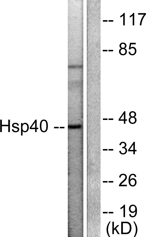
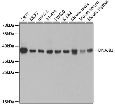
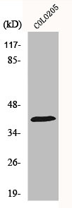
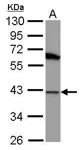
Figure 1. Western blot analysis of Hsp40/DNAJB1 using anti-Hsp40/DNAJB1 antibody (A03100-1). Electrophoresis was performed on a 5-20% SDS-PAGE gel at 70V (Stacking gel) / 90V (Resolving gel) for 2-3 hours. The sample well of each lane was loaded with 30 ug of sample under reducing conditions. Lane 1: human U251 whole cell lysates, Lane 2: human HepG2 whole cell lysates, Lane 3: human PC-3 whole cell lysates, Lane 4: human A549 whole cell lysates, Lane 5: rat testis tissue lysates, Lane 6: mouse testis tissue lysates. After electrophoresis, proteins were transferred to a nitrocellulose membrane at 150 mA for 50-90 minutes. Blocked the membrane with 5% non-fat milk/TBS for 1.5 hour at RT. The membrane was incubated with rabbit anti-Hsp40/DNAJB1 antigen affinity purified polyclonal antibody (Catalog # A03100-1) at 0.25 microg/mL overnight at 4°C, then washed with TBS-0.1%Tween 3 times with 5 minutes each and probed with a goat anti-rabbit IgG-HRP secondary antibody at a dilution of 1:5000 for 1.5 hour at RT. The signal is developed using an Enhanced Chemiluminescent detection (ECL) kit (Catalog # EK1002) with Tanon 5200 system. A specific band was detected for Hsp40/DNAJB1 at approximately 38 kDa. The expected band size for Hsp40/DNAJB1 is at 38 kDa.
. Hsp40/DNAJB1 was detected in a paraffin-embedded section of human gastric signet ring cell carcinoma tissue. Heat mediated antigen retrieval was performed in EDTA buffer (pH 8.0, epitope retrieval solution). The tissue section was blocked with 10% goat serum. The tissue section was then incubated with 2 microg/ml rabbit anti-Hsp40/DNAJB1 Antibody (A03100-1) overnight at 4°C. Biotinylated goat anti-rabbit IgG was used as secondary antibody and incubated for 30 minutes at 37°C. The tissue section was developed using Strepavidin-Biotin-Complex (SABC) (Catalog # SA1022) with DAB as the chromogen.")
. Hsp40/DNAJB1 was detected in an immunocytochemical section of A549 cells. Enzyme antigen retrieval was performed using IHC enzyme antigen retrieval reagent (AR0022) for 15 mins. The cells were blocked with 10% goat serum. And then incubated with 5 microg/mL rabbit anti-Hsp40/DNAJB1 Antibody (A03100-1) overnight at 4°C. DyLight®488 Conjugated Goat Anti-Rabbit IgG (BA1127) was used as secondary antibody at 1:100 dilution and incubated for 30 minutes at 37°C. The section was counterstained with DAPI. Visualize using a fluorescence microscope and filter sets appropriate for the label used.")
. Overlay histogram showing C6 cells stained with A03100-1 (Blue line). To facilitate intracellular staining, cells were fixed with 4% paraformaldehyde and permeabilized with permeabilization buffer. The cells were blocked with 10% normal goat serum. And then incubated with rabbit anti-Hsp40/DNAJB1 Antibody (A03100-1, 1 microg/1x106 cells) for 30 min at 20°C. DyLight®488 conjugated goat anti-rabbit IgG (BA1127, 5-10 microg/1x106 cells) was used as secondary antibody for 30 minutes at 20°C. Isotype control antibody (Green line) was rabbit IgG (1 microg/1x106) used under the same conditions. Unlabelled sample without incubation with primary antibody and secondary antibody (Red line) was used as a blank control.")
Figure 1. Western blot analysis of Hsp40/DNAJB1 using anti-Hsp40/DNAJB1 antibody (A03100-1). Electrophoresis was performed on a 5-20% SDS-PAGE gel at 70V (Stacking gel) / 90V (Resolving gel) for 2-3 hours. The sample well of each lane was loaded with 30 ug of sample under reducing conditions. Lane 1: human U251 whole cell lysates, Lane 2: human HepG2 whole cell lysates, Lane 3: human PC-3 whole cell lysates, Lane 4: human A549 whole cell lysates, Lane 5: rat testis tissue lysates, Lane 6: mouse testis tissue lysates. After electrophoresis, proteins were transferred to a nitrocellulose membrane at 150 mA for 50-90 minutes. Blocked the membrane with 5% non-fat milk/TBS for 1.5 hour at RT. The membrane was incubated with rabbit anti-Hsp40/DNAJB1 antigen affinity purified polyclonal antibody (Catalog # A03100-1) at 0.25 microg/mL overnight at 4°C, then washed with TBS-0.1%Tween 3 times with 5 minutes each and probed with a goat anti-rabbit IgG-HRP secondary antibody at a dilution of 1:5000 for 1.5 hour at RT. The signal is developed using an Enhanced Chemiluminescent detection (ECL) kit (Catalog # EK1002) with Tanon 5200 system. A specific band was detected for Hsp40/DNAJB1 at approximately 38 kDa. The expected band size for Hsp40/DNAJB1 is at 38 kDa.
Anti-Hsp40/DNAJB1 Antibody Picoband(r)
A03100-1-CARRIER-FREE
ApplicationsFlow Cytometry, ImmunoFluorescence, Western Blot, ImmunoCytoChemistry, ImmunoHistoChemistry
Product group Antibodies
ReactivityHuman, Mouse, Rat
TargetDNAJB1
Overview
- SupplierBoster Bio
- Product NameAnti-Hsp40/DNAJB1 Antibody Picoband(r)
- Delivery Days Customer9
- ApplicationsFlow Cytometry, ImmunoFluorescence, Western Blot, ImmunoCytoChemistry, ImmunoHistoChemistry
- CertificationResearch Use Only
- ClonalityPolyclonal
- Concentration500 ug/ml
- Gene ID3337
- Target nameDNAJB1
- Target descriptionDnaJ heat shock protein family (Hsp40) member B1
- Target synonymsHSPF1, Hdj1, Hsp40, RSPH16B, Sis1, dnaJ homolog subfamily B member 1, DnaJ (Hsp40) homolog, subfamily B, member 1, dnaJ protein homolog 1, heat shock 40 kDa protein 1, human DnaJ protein 1, radial spoke 16 homolog B
- HostRabbit
- IsotypeIgG
- Protein IDP25685
- Protein NameDnaJ homolog subfamily B member 1
- Scientific DescriptionBoster Bio Anti-Hsp40/DNAJB1 Antibody Picoband® catalog # A03100-1. Tested in Flow Cytometry, IF, IHC, ICC, WB applications. This antibody reacts with Human, Mouse, Rat. The brand Picoband indicates this is a premium antibody that guarantees superior quality, high affinity, and strong signals with minimal background in Western blot applications. Only our best-performing antibodies are designated as Picoband, ensuring unmatched performance.
- ReactivityHuman, Mouse, Rat
- Storage Instruction-20°C,2°C to 8°C
- UNSPSC12352203
Related products
Product group Antibodies
Anti-HSP40 AntibodyA95221
ApplicationsImmunoFluorescence, Western Blot, ELISA, ImmunoHistoChemistry
ReactivityHuman, Mouse, Rat
- SizePrice
Product group Antibodies
Anti-DNAJB1 Antibody144-05504
ApplicationsImmunoFluorescence, Western Blot, ImmunoHistoChemistry
ReactivityHuman, Mouse
TargetDNAJB1
- SizePrice
Product group Antibodies
DNAJB1 Polyclonal AntibodyBS-55055R
ApplicationsImmunoFluorescence, Western Blot, ImmunoCytoChemistry
ReactivityHuman, Mouse, Rat
TargetDNAJB1
- SizePrice
Product group Antibodies
DNAJB1 AntibodyCSB-PA002989
ApplicationsImmunoFluorescence, Western Blot, ELISA, ImmunoHistoChemistry
ReactivityHuman, Mouse, Rat
TargetDNAJB1
- SizePrice
Product group Antibodies
DNAJB1 Polyclonal AntibodyCAC14054
ApplicationsWestern Blot, ELISA, ImmunoHistoChemistry
ReactivityMouse
TargetDNAJB1
- SizePrice
Product group Antibodies

HSP40 AntibodyLS-C404589
ApplicationsWestern Blot, ELISA, ImmunoHistoChemistry
ReactivityHuman, Mouse
- SizePrice
Product group Antibodies
Hsp40 antibody [N1C1]GTX102049
ApplicationsWestern Blot
ReactivityHuman
TargetDNAJB1
- SizePrice

Product group Antibodies
Anti-DNAJB1 AntibodyHPA063247
ApplicationsImmunoCytoChemistry, ImmunoHistoChemistry
ReactivityHuman
TargetDNAJB1
- SizePrice
Product group Antibodies
ApplicationsImmunoFluorescence, Western Blot, ELISA, ImmunoCytoChemistry
ReactivityHuman
TargetDNAJB1
- SizePrice