


Immunofluorescent staining of human cell line U-2 OS shows localization to cytosol & the Golgi apparatus.
Immunofluorescent staining of human cell line U-2 OS shows localization to cytosol & the Golgi apparatus.
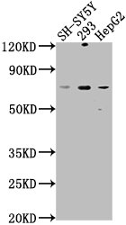
Anti-HSPA12A Antibody
HPA073244
ApplicationsImmunoCytoChemistry
Product group Antibodies
ReactivityHuman
TargetHSPA12A
Overview
- SupplierAtlas Antibodies
- Product NameAnti-HSPA12A Antibody
- Delivery Days Customer4
- ApplicationsImmunoCytoChemistry
- CertificationResearch Use Only
- ClonalityPolyclonal
- ConjugateUnconjugated
- Gene ID259217
- Target nameHSPA12A
- Target descriptionheat shock protein family A (Hsp70) member 12A
- Target synonymsheat shock 70 kDa protein 12A, heat shock 70kD protein 12A, heat shock 70kDa protein 12A, heat shock protein family A member 12A
- HostRabbit
- IsotypeIgG
- Protein IDO43301
- Protein NameHeat shock 70 kDa protein 12A
- Scientific DescriptionRecombinant Protein Epitope Signature Tag (PrEST) antigen sequence
- ReactivityHuman
- Storage Instruction-20°C,2°C to 8°C
- UNSPSC41116161
Datasheet
MSDS
Related products
Product group Antibodies
HSPA12A AntibodyCSB-PA010817LA01HU
ApplicationsWestern Blot, ELISA
ReactivityHuman
TargetHSPA12A
- SizePrice
Product group Antibodies
HSPA12A Polyclonal AntibodyCAC15812
ApplicationsWestern Blot, ELISA
TargetHSPA12A
- SizePrice

Product group Antibodies
Anti-HSPA12A Antibody Picoband(r)A13632-1-CARRIER-FREE
ApplicationsImmunoFluorescence, Western Blot, ELISA, ImmunoCytoChemistry
ReactivityHuman, Mouse, Rat
TargetHSPA12A
- SizePrice
Product group Antibodies

HSPA12A AntibodyLS-C779308
ApplicationsWestern Blot, ELISA
ReactivityHuman, Mouse
TargetHSPA12A
- SizePrice

Product group Antibodies
Anti-HSPA12A AntibodyHPA011273
ApplicationsWestern Blot, ImmunoHistoChemistry
ReactivityHuman
TargetHSPA12A
- SizePrice
Product group Antibodies
HSPA12A Recombinant AntibodyBSM-61137R
ApplicationsImmunoFluorescence, ImmunoPrecipitation, Western Blot, ImmunoCytoChemistry, ImmunoHistoChemistry, ImmunoHistoChemistry Frozen, ImmunoHistoChemistry Paraffin
TargetHSPA12A
- SizePrice