

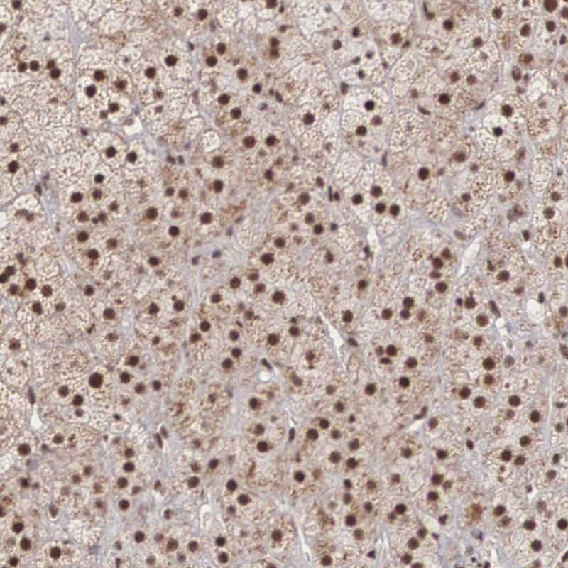
Immunohistochemical staining of human adrenal gland shows strong nuclear positivity in glandular cells.
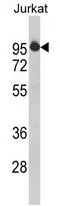
![Lane 1: Marker [kDa] 250, 130, 100, 70, 55, 35, 25, 15, 10. Lane 2: Human Adrenal Gland tissue](https://atlasantibodies.s3.amazonaws.com/images/wb/hpa000504-wb-1.jpg "Lane 1: Marker [kDa] 250, 130, 100, 70, 55, 35, 25, 15, 10. Lane 2: Human Adrenal Gland tissue")
Immunohistochemical staining of human adrenal gland shows strong nuclear positivity in glandular cells.
Anti-HTATSF1 Antibody
HPA000504
ApplicationsWestern Blot, ChIP Chromatin ImmunoPrecipitation, ImmunoCytoChemistry, ImmunoHistoChemistry
Product group Antibodies
ReactivityHuman
TargetHTATSF1
Overview
- SupplierAtlas Antibodies
- Product NameAnti-HTATSF1 Antibody
- Delivery Days Customer4
- ApplicationsWestern Blot, ChIP Chromatin ImmunoPrecipitation, ImmunoCytoChemistry, ImmunoHistoChemistry
- CertificationResearch Use Only
- ClonalityPolyclonal
- ConjugateUnconjugated
- Gene ID27336
- Target nameHTATSF1
- Target descriptionHIV-1 Tat specific factor 1
- Target synonymsTAT-SF1, TATSF1, dJ196E23.2, 17S U2 SnRNP complex component HTATSF1, HIV TAT specific factor 1, HIV Tat-specific factor 1, cofactor required for Tat activation of HIV-1 transcription
- HostRabbit
- IsotypeIgG
- Protein IDO43719
- Protein Name17S U2 SnRNP complex component HTATSF1
- Scientific DescriptionRecombinant Protein Epitope Signature Tag (PrEST) antigen sequence
- ReactivityHuman
- Storage Instruction-20°C,2°C to 8°C
- UNSPSC41116161
Datasheet
MSDS
Related products
Product group Antibodies
HTATSF1 AntibodyCSB-PA010872GA01HU
ApplicationsImmunoFluorescence, Western Blot, ELISA, ImmunoHistoChemistry
ReactivityHuman, Mouse, Rat
TargetHTATSF1
- SizePrice

Product group Antibodies
Anti-HTATSF1 Antibody Picoband(r)A07890-2-CARRIER-FREE
ApplicationsFlow Cytometry, ImmunoFluorescence, Western Blot, ELISA, ImmunoHistoChemistry
ReactivityHuman, Mouse, Rat
TargetHTATSF1
- SizePrice
Product group Antibodies

HTATSF1 / TAT-SF1 AntibodyLS-C748467
ApplicationsWestern Blot
ReactivityHuman, Mouse, Rat
TargetHTATSF1
- SizePrice
Product group Antibodies
References

Goat anti-HTATSF1EB07999
ApplicationsWestern Blot, ELISA
ReactivityCanine, Human, Rat
TargetHTATSF1
- SizePrice
Product group Antibodies
HTATSF1 antibody, N-termGTX81789
ApplicationsWestern Blot, ImmunoHistoChemistry, ImmunoHistoChemistry Paraffin
ReactivityHuman
TargetHTATSF1
- SizePrice
Product group Antibodies
ApplicationsImmunoFluorescence, Western Blot
ReactivityHuman, Mouse
TargetHTATSF1
- SizePrice