

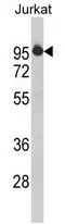
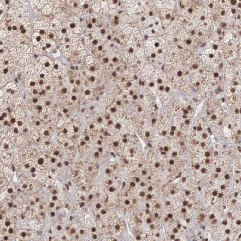
Anti-HTATSF1 Antibody
CAB5977
ApplicationsWestern Blot, ELISA, ImmunoHistoChemistry, ImmunoHistoChemistry Paraffin
Product group Antibodies
ReactivityHuman
TargetHTATSF1
Overview
- SupplierAssay Genie
- Product NameAnti-HTATSF1 Antibody
- Delivery Days Customer9
- ApplicationsWestern Blot, ELISA, ImmunoHistoChemistry, ImmunoHistoChemistry Paraffin
- Applications SupplierWB,IHC,IF
- CertificationResearch Use Only
- ClonalityPolyclonal
- Clone IDNot applicable
- ConjugateUnconjugated
- Gene ID27336
- Target nameHTATSF1
- Target descriptionHIV-1 Tat specific factor 1
- Target synonymsTAT-SF1, TATSF1, dJ196E23.2, 17S U2 SnRNP complex component HTATSF1, HIV TAT specific factor 1, HIV Tat-specific factor 1, cofactor required for Tat activation of HIV-1 transcription
- HostRabbit
- Protein IDO43719
- Protein Name17S U2 SnRNP complex component HTATSF1
- Scientific DescriptionHTATSF1 Antibody is a premium polyclonal antibody that offers outstanding performance and reliability for demanding research applications. Rigorously validated for WB, IHC-P, ELISA, this antibody ensures consistent, reproducible results across multiple experimental platforms. Demonstrates excellent reactivity with Human,Mouse,Rat samples, providing researchers with confidence in cross-species compatibility. Conveniently packaged in 100microL format to meet your experimental needs. For optimal performance, store at -20°C and maintains stability for 12 months. Backed by rigorous quality control testing to ensure superior performance in your critical research applications.
- Shelf life instruction12 months
- SourceRabbit
- ReactivityHuman
- Reactivity SupplierHuman,Mouse,Rat
- Storage Instruction-20°C
- UNSPSC41116161
Related products

Product group Antibodies
Anti-HTATSF1 Antibody Picoband(r)A07890-2-CARRIER-FREE
ApplicationsFlow Cytometry, ImmunoFluorescence, Western Blot, ELISA, ImmunoHistoChemistry
ReactivityHuman, Mouse, Rat
TargetHTATSF1
- SizePrice
Product group Antibodies

HTATSF1 / TAT-SF1 AntibodyLS-C748467
ApplicationsWestern Blot
ReactivityHuman, Mouse, Rat
TargetHTATSF1
- SizePrice
Product group Antibodies
ApplicationsImmunoFluorescence, Western Blot
ReactivityHuman, Mouse
TargetHTATSF1
- SizePrice
Product group Antibodies
HTATSF1 AntibodyCSB-PA010872GA01HU
ApplicationsImmunoFluorescence, Western Blot, ELISA, ImmunoHistoChemistry
ReactivityHuman, Mouse, Rat
TargetHTATSF1
- SizePrice
Product group Antibodies
References

Goat anti-HTATSF1EB07999
ApplicationsWestern Blot, ELISA
ReactivityCanine, Human, Rat
TargetHTATSF1
- SizePrice
Product group Antibodies
HTATSF1 antibody, N-termGTX81789
ApplicationsWestern Blot, ImmunoHistoChemistry, ImmunoHistoChemistry Paraffin
ReactivityHuman
TargetHTATSF1
- SizePrice
Product group Antibodies
Anti-HTATSF1 AntibodyHPA000504
ApplicationsWestern Blot, ChIP Chromatin ImmunoPrecipitation, ImmunoCytoChemistry, ImmunoHistoChemistry
ReactivityHuman
TargetHTATSF1
- SizePrice