


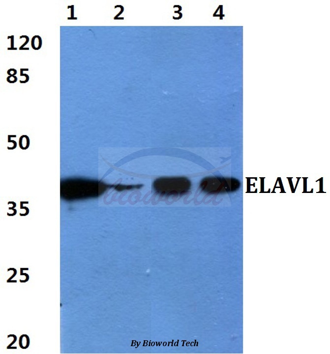
Figure 1. Western blot analysis of ELAVL1 using anti-ELAVL1 antibody (M00736). Electrophoresis was performed on a 5-20% SDS-PAGE gel at 70V (Stacking gel) / 90V (Resolving gel) for 2-3 hours. The sample well of each lane was loaded with 30 ug of sample under reducing conditions. Lane 1: human Jurkat whole cell lysates, Lane 2: human Hela whole cell lysates, Lane 3: human MOLT-4 whole cell lysates, Lane 4: human HepG2 whole cell lysates, Lane 5: rat heart tissue lysates, Lane 6: rat spleen tissue lysates, Lane 7: mouse heart tissue lysates, Lane 8: mouse spleen tissue lysates. After electrophoresis, proteins were transferred to a nitrocellulose membrane at 150 mA for 50-90 minutes. Blocked the membrane with 5% non-fat milk/TBS for 1.5 hour at RT. The membrane was incubated with rabbit anti-ELAVL1 antigen affinity purified monoclonal antibody (Catalog # M00736) at 1:500 overnight at 4°C, then washed with TBS-0.1%Tween 3 times with 5 minutes each and probed with a goat anti-rabbit IgG-HRP secondary antibody at a dilution of 1:500 for 1.5 hour at RT. The signal is developed using an Enhanced Chemiluminescent detection (ECL) kit (Catalog # EK1002) with Tanon 5200 system. A specific band was detected for ELAVL1 at approximately 36 kDa. The expected band size for ELAVL1 is at 36 kDa.
Figure 1. Western blot analysis of ELAVL1 using anti-ELAVL1 antibody (M00736). Electrophoresis was performed on a 5-20% SDS-PAGE gel at 70V (Stacking gel) / 90V (Resolving gel) for 2-3 hours. The sample well of each lane was loaded with 30 ug of sample under reducing conditions. Lane 1: human Jurkat whole cell lysates, Lane 2: human Hela whole cell lysates, Lane 3: human MOLT-4 whole cell lysates, Lane 4: human HepG2 whole cell lysates, Lane 5: rat heart tissue lysates, Lane 6: rat spleen tissue lysates, Lane 7: mouse heart tissue lysates, Lane 8: mouse spleen tissue lysates. After electrophoresis, proteins were transferred to a nitrocellulose membrane at 150 mA for 50-90 minutes. Blocked the membrane with 5% non-fat milk/TBS for 1.5 hour at RT. The membrane was incubated with rabbit anti-ELAVL1 antigen affinity purified monoclonal antibody (Catalog # M00736) at 1:500 overnight at 4°C, then washed with TBS-0.1%Tween 3 times with 5 minutes each and probed with a goat anti-rabbit IgG-HRP secondary antibody at a dilution of 1:500 for 1.5 hour at RT. The signal is developed using an Enhanced Chemiluminescent detection (ECL) kit (Catalog # EK1002) with Tanon 5200 system. A specific band was detected for ELAVL1 at approximately 36 kDa. The expected band size for ELAVL1 is at 36 kDa.
Anti-HuR / ELAVL1 Monoclonal Antibody
M00736
ApplicationsFlow Cytometry, ImmunoFluorescence, ImmunoPrecipitation, Western Blot, ImmunoCytoChemistry, ImmunoHistoChemistry
Product group Antibodies
ReactivityHuman, Mouse, Rat
TargetELAVL1
Overview
- SupplierBoster Bio
- Product NameAnti-HuR / ELAVL1 Monoclonal Antibody
- Delivery Days Customer9
- ApplicationsFlow Cytometry, ImmunoFluorescence, ImmunoPrecipitation, Western Blot, ImmunoCytoChemistry, ImmunoHistoChemistry
- CertificationResearch Use Only
- ClonalityMonoclonal
- Clone IDADHI-5
- Gene ID1994
- Target nameELAVL1
- Target descriptionELAV like RNA binding protein 1
- Target synonymsELAV1, HUR, Hua, MelG, ELAV-like protein 1, ELAV (embryonic lethal, abnormal vision, Drosophila)-like 1 (Hu antigen R), Hu antigen R, Human antigen R, embryonic lethal, abnormal vision, drosophila, homolog-like 1, hu-antigen R
- HostRabbit
- IsotypeIgG
- Protein IDQ15717
- Protein NameELAV-like protein 1
- Scientific DescriptionBoster Bio Anti-HuR / ELAVL1 Monoclonal Antibody catalog # M00736. Tested in WB, IHC, ICC/IF, IP, Flow Cytometry applications. This antibody reacts with Human, Mouse, Rat.
- ReactivityHuman, Mouse, Rat
- Storage Instruction-20°C
- UNSPSC12352203
Datasheet
MSDS
Related products
Product group Antibodies
Anti-ELAVL1 AntibodyA28604
ApplicationsWestern Blot
ReactivityHuman, Mouse, Rat
- SizePrice
Product group Antibodies
Anti-ELAVL1 Antibody144-01608
ApplicationsImmunoFluorescence, Western Blot, ImmunoHistoChemistry
ReactivityHuman, Mouse, Rat
TargetELAVL1
- SizePrice
Product group Antibodies
ELAVL1 Recombinant Antibody, AbBy Fluor-350 ConjugatedBSM-61688R-BF350
ApplicationsFlow Cytometry, ImmunoFluorescence, Western Blot
ReactivityHuman, Mouse, Rat
TargetELAVL1
- SizePrice
Product group Antibodies
ELAVL1 AntibodyCSB-PA104194
ApplicationsWestern Blot, ELISA, ImmunoHistoChemistry
ReactivityHuman, Mouse
TargetELAVL1
- SizePrice
Product group Antibodies
Elavl1 Polyclonal AntibodyCAC07564
ApplicationsWestern Blot, ELISA
TargetELAVL1
- SizePrice
Product group Antibodies

ELAVL1 / HUR AntibodyLS-C400726
ApplicationsWestern Blot, ELISA, ImmunoHistoChemistry
ReactivityHuman, Mouse
TargetELAVL1
- SizePrice
Product group Antibodies
ELAVL1 / HuR antibodyGTX134821
ApplicationsImmunoFluorescence, Western Blot, ImmunoCytoChemistry
ReactivityHuman, Mouse
TargetELAVL1
- SizePrice
Product group Antibodies
Anti-ELAVL1 AntibodyHPA046298
ApplicationsImmunoHistoChemistry
ReactivityHuman
TargetELAVL1
- SizePrice