

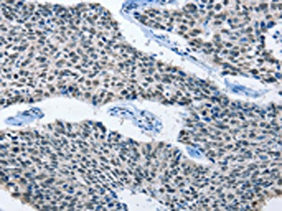
The image on the left is immunohistochemistry of paraffin-embedded Human lung cancer tissue using CSB-PA104194(ELAVL1 Antibody) at dilution 1/50, on the right is treated with fusion protein. (Original magnification: x200)
 at dilution 1/50, on the right is treated with fusion protein. (Original magnification: x200)")
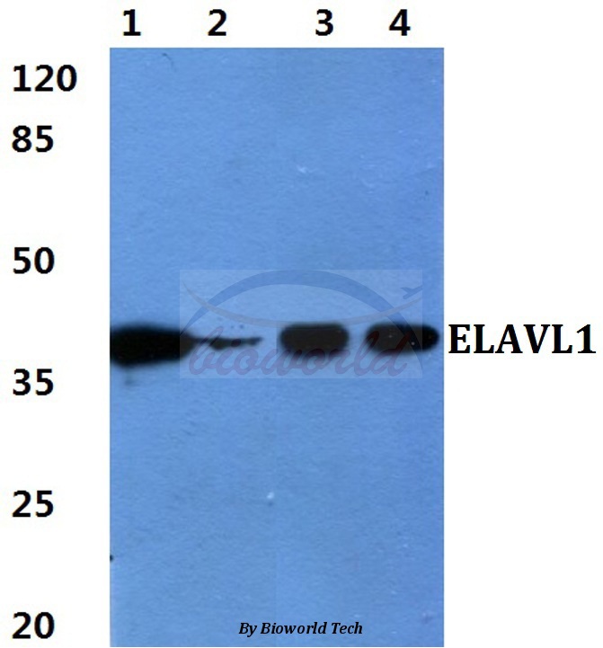
 at dilution 1/800, Secondary antibody: Goat anti rabbit IgG at 1/8000 dilution, Exposure time: 5 seconds")
The image on the left is immunohistochemistry of paraffin-embedded Human lung cancer tissue using CSB-PA104194(ELAVL1 Antibody) at dilution 1/50, on the right is treated with fusion protein. (Original magnification: x200)
ELAVL1 Antibody
CSB-PA104194
ApplicationsWestern Blot, ELISA, ImmunoHistoChemistry
Product group Antibodies
ReactivityHuman, Mouse
TargetELAVL1
Overview
- SupplierCusabio
- Product NameELAVL1 Antibody
- Delivery Days Customer20
- ApplicationsWestern Blot, ELISA, ImmunoHistoChemistry
- CertificationResearch Use Only
- ClonalityPolyclonal
- ConjugateUnconjugated
- Gene ID1994
- Target nameELAVL1
- Target descriptionELAV like RNA binding protein 1
- Target synonymsELAV1, HUR, Hua, MelG, ELAV-like protein 1, ELAV (embryonic lethal, abnormal vision, Drosophila)-like 1 (Hu antigen R), Hu antigen R, Human antigen R, embryonic lethal, abnormal vision, drosophila, homolog-like 1, hu-antigen R
- HostRabbit
- IsotypeIgG
- Protein IDQ15717
- Protein NameELAV-like protein 1
- Scientific DescriptionThe protein encoded by this gene is a member of the ELAVL family of RNA-binding proteins that contain several RNA recognition motifs, and selectively bind AU-rich elements (AREs) found in the 3 untranslated regions of mRNAs. AREs signal degradation of mRNAs as a means to regulate gene expression, thus by binding AREs, the ELAVL family of proteins play a role in stabilizing ARE-containing mRNAs. This gene has been implicated in a variety of biological processes and has been linked to a number of diseases, including cancer. It is highly expressed in many cancers, and could be potentially useful in cancer diagnosis, prognosis, and therapy.
- ReactivityHuman, Mouse
- Storage Instruction-20°C or -80°C
- UNSPSC41116161
Related products
Product group Antibodies
Anti-ELAVL1 AntibodyA28604
ApplicationsWestern Blot
ReactivityHuman, Mouse, Rat
- SizePrice
Product group Antibodies
Anti-ELAVL1 Antibody144-01608
ApplicationsImmunoFluorescence, Western Blot, ImmunoHistoChemistry
ReactivityHuman, Mouse, Rat
TargetELAVL1
- SizePrice

Product group Antibodies
ApplicationsFlow Cytometry, ImmunoFluorescence, ImmunoPrecipitation, Western Blot, ImmunoCytoChemistry, ImmunoHistoChemistry
ReactivityHuman, Mouse, Rat
TargetELAVL1
- SizePrice
Product group Antibodies
ELAVL1 Recombinant Antibody, AbBy Fluor-350 ConjugatedBSM-61688R-BF350
ApplicationsFlow Cytometry, ImmunoFluorescence, Western Blot
ReactivityHuman, Mouse, Rat
TargetELAVL1
- SizePrice
Product group Antibodies
Elavl1 Polyclonal AntibodyCAC07564
ApplicationsWestern Blot, ELISA
TargetELAVL1
- SizePrice
Product group Antibodies

ELAVL1 / HUR AntibodyLS-C400726
ApplicationsWestern Blot, ELISA, ImmunoHistoChemistry
ReactivityHuman, Mouse
TargetELAVL1
- SizePrice
Product group Antibodies
ELAVL1 / HuR antibodyGTX134821
ApplicationsImmunoFluorescence, Western Blot, ImmunoCytoChemistry
ReactivityHuman, Mouse
TargetELAVL1
- SizePrice
Product group Antibodies
Anti-ELAVL1 AntibodyHPA046298
ApplicationsImmunoHistoChemistry
ReactivityHuman
TargetELAVL1
- SizePrice