

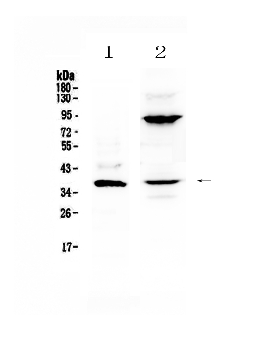
Figure 1. Western blot analysis of IL12B using anti-IL12B antibody (A01152-4). Electrophoresis was performed on a 5-20% SDS-PAGE gel at 70V (Stacking gel) / 90V (Resolving gel) for 2-3 hours. The sample well of each lane was loaded with 50ug of sample under reducing conditions. Lane 1: mouse NIH3T3 cell lysates, Lane 2: human A549 cell lysates. After Electrophoresis, proteins were transferred to a Nitrocellulose membrane at 150mA for 50-90 minutes. Blocked the membrane with 5% Non-fat Milk/ TBS for 1.5 hour at RT. The membrane was incubated with rabbit anti-IL12B antigen affinity purified polyclonal antibody (Catalog # A01152-4) at 0.5 microg/mL overnight at 4°C, then washed with TBS-0.1%Tween 3 times with 5 minutes each and probed with a goat anti-rabbit IgG-HRP secondary antibody at a dilution of 1:10000 for 1.5 hour at RT. The signal is developed using an Enhanced Chemiluminescent detection (ECL) kit (Catalog # EK1002) with Tanon 5200 system. A specific band was detected for IL12B at approximately 37KD. The expected band size for IL12B is at 37KD.
Figure 1. Western blot analysis of IL12B using anti-IL12B antibody (A01152-4). Electrophoresis was performed on a 5-20% SDS-PAGE gel at 70V (Stacking gel) / 90V (Resolving gel) for 2-3 hours. The sample well of each lane was loaded with 50ug of sample under reducing conditions. Lane 1: mouse NIH3T3 cell lysates, Lane 2: human A549 cell lysates. After Electrophoresis, proteins were transferred to a Nitrocellulose membrane at 150mA for 50-90 minutes. Blocked the membrane with 5% Non-fat Milk/ TBS for 1.5 hour at RT. The membrane was incubated with rabbit anti-IL12B antigen affinity purified polyclonal antibody (Catalog # A01152-4) at 0.5 microg/mL overnight at 4°C, then washed with TBS-0.1%Tween 3 times with 5 minutes each and probed with a goat anti-rabbit IgG-HRP secondary antibody at a dilution of 1:10000 for 1.5 hour at RT. The signal is developed using an Enhanced Chemiluminescent detection (ECL) kit (Catalog # EK1002) with Tanon 5200 system. A specific band was detected for IL12B at approximately 37KD. The expected band size for IL12B is at 37KD.
Anti-IL12B Antibody Picoband(r)
A01152-4-CARRIER-FREE
ApplicationsWestern Blot, ELISA
Product group Antibodies
ReactivityHuman, Mouse
TargetIL12B
Overview
- SupplierBoster Bio
- Product NameAnti-IL12B Antibody Picoband(r)
- Delivery Days Customer9
- ApplicationsWestern Blot, ELISA
- CertificationResearch Use Only
- ClonalityPolyclonal
- Concentration500 ug/ml
- Gene ID3593
- Target nameIL12B
- Target descriptioninterleukin 12B
- Target synonymsCLMF, CLMF2, IL-12B, IMD28, IMD29, NKSF, NKSF2, interleukin-12 subunit beta, CLMF p40, IL-12 subunit p40, IL12, subunit p40, NK cell stimulatory factor chain 2, cytotoxic lymphocyte maturation factor 40 kDa subunit, interleukin 12, p40, interleukin 12B (natural killer cell stimulatory factor 2, cytotoxic lymphocyte maturation factor 2, p40), interleukin-12 beta chain, natural killer cell stimulatory factor, 40 kD subunit
- HostRabbit
- IsotypeIgG
- Protein IDP29460
- Protein NameInterleukin-12 subunit beta
- Scientific DescriptionBoster Bio Anti-IL12B Antibody Picoband® catalog # A01152-4. Tested in ELISA, WB applications. This antibody reacts with Human, Mouse. The brand Picoband indicates this is a premium antibody that guarantees superior quality, high affinity, and strong signals with minimal background in Western blot applications. Only our best-performing antibodies are designated as Picoband, ensuring unmatched performance.
- ReactivityHuman, Mouse
- Storage Instruction-20°C,2°C to 8°C
- UNSPSC12352203
Related products
Product group Antibodies

Anti-IL-12/23 [ABT-874 (Briakinumab)]Ab00722-10.0
ApplicationsNeutralisation/Blocking
ReactivityHuman
TargetIL12B
- SizePrice
Product group Antibodies
Anti-IL-12B AntibodyA100523
ApplicationsWestern Blot, ELISA
ReactivityHuman
- SizePrice
Product group Antibodies
Anti-IL12 beta Antibody130-10269
ApplicationsWestern Blot, ELISA
ReactivityHuman
TargetIL12B
- SizePrice
Product group Antibodies
IL12B Polyclonal AntibodyBS-10641R
ApplicationsImmunoFluorescence, Western Blot, ELISA, ImmunoCytoChemistry, ImmunoHistoChemistry, ImmunoHistoChemistry Frozen, ImmunoHistoChemistry Paraffin
TargetIL12B
- SizePrice
Product group Antibodies
IL12B AntibodyCSB-PA009590
ApplicationsWestern Blot, ELISA
ReactivityHuman
TargetIL12B
- SizePrice
Product group Antibodies

Goat anti-IL12B / IL12p40EB07862
ApplicationsWestern Blot, ELISA
ReactivityHuman
TargetIL12B
- SizePrice
Product group Antibodies
Il12B Polyclonal AntibodyCAC07032
ApplicationsWestern Blot, ELISA, ImmunoHistoChemistry
ReactivityMouse
TargetIL12B
- SizePrice
Product group Antibodies

ApplicationsWestern Blot, ELISA
ReactivityHuman
TargetIL12B
- SizePrice
Product group Antibodies
IL12B / IL12 p40 antibodyGTX114135
ApplicationsWestern Blot
ReactivityHuman
TargetIL12B
- SizePrice