


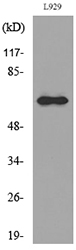
Figure 1. Western blot analysis of IL2 Receptor beta/p75/IL2RB using anti-IL2 Receptor beta/p75/IL2RB antibody (A03401-3). Electrophoresis was performed on a 5-20% SDS-PAGE gel at 70V (Stacking gel) / 90V (Resolving gel) for 2-3 hours. The sample well of each lane was loaded with 30ug of sample under reducing conditions. Lane 1: human Hela whole cell lysates, Lane 2: human Hek293 whole cell lysates, Lane 3: human K562 whole cell lysates, Lane 4: human THP-1 whole cell lysates, Lane 5: rat PC-12 whole cell lysates, Lane 6: mouse intestine tissue lysates, Lane 7: mouse RAW264.7 whole cell lysates. After Electrophoresis, proteins were transferred to a Nitrocellulose membrane at 150mA for 50-90 minutes. Blocked the membrane with 5% Non-fat Milk/ TBS for 1.5 hour at RT. The membrane was incubated with rabbit anti-IL2 Receptor beta/p75/IL2RB antigen affinity purified polyclonal antibody (Catalog # A03401-3) at 0.5 microg/mL overnight at 4°C, then washed with TBS-0.1%Tween 3 times with 5 minutes each and probed with a goat anti-rabbit IgG-HRP secondary antibody at a dilution of 1:5000 for 1.5 hour at RT. The signal is developed using an Enhanced Chemiluminescent detection (ECL) kit (Catalog # EK1002) with Tanon 5200 system. A specific band was detected for IL2 Receptor beta/p75/IL2RB at approximately 75KD. The expected band size for IL2 Receptor beta/p75/IL2RB is at 75KD.
. IL2 Receptor beta/p75/IL2RB was detected in immunocytochemical section of A549 cells. Enzyme antigen retrieval was performed using IHC enzyme antigen retrieval reagent (AR0022) for 15 mins. The cells were blocked with 10% goat serum. And then incubated with 5microg/mL rabbit anti- IL2 Receptor beta/p75/IL2RB Antibody (A03401-3) overnight at 4°C. DyLight®488 Conjugated Goat Anti-Rabbit IgG (BA1127) was used as secondary antibody at 1:100 dilution and incubated for 30 minutes at 37°C. The section was counterstained with DAPI. Visualize using a fluorescence microscope and filter sets appropriate for the label used.")
. Overlay histogram showing U20S cells stained with A03401-3 (Blue line). The cells were fixed with 4% paraformaldehyde and blocked with 10% normal goat serum. And then incubated with rabbit anti-IL2 Receptor beta/p75/IL2RB Antibody (A03401-3, 1microg/1x106 cells) for 30 min at 20°C. DyLight®488 conjugated goat anti-rabbit IgG (BA1127, 5-10microg/1x106 cells) was used as secondary antibody for 30 minutes at 20°C. Isotype control antibody (Green line) was rabbit IgG (1microg/1x106) used under the same conditions. Unlabelled sample without incubation with primary antibody and secondary antibody (Red line) was used as a blank control.")
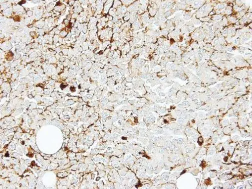
. IL2 Receptor beta/p75/IL2RB was detected in a paraffin-embedded section of human appendicitis tissue. Heat mediated antigen retrieval was performed in EDTA buffer (pH 8.0, epitope retrieval solution). The tissue section was blocked with 10% goat serum. The tissue section was then incubated with 2 microg/ml rabbit anti-IL2 Receptor beta/p75/IL2RB Antibody (A03401-3) overnight at 4°C. Biotinylated goat anti-rabbit IgG was used as secondary antibody and incubated for 30 minutes at 37°C. The tissue section was developed using Strepavidin-Biotin-Complex (SABC) (Catalog # SA1022) with DAB as the chromogen.")
Figure 1. Western blot analysis of IL2 Receptor beta/p75/IL2RB using anti-IL2 Receptor beta/p75/IL2RB antibody (A03401-3). Electrophoresis was performed on a 5-20% SDS-PAGE gel at 70V (Stacking gel) / 90V (Resolving gel) for 2-3 hours. The sample well of each lane was loaded with 30ug of sample under reducing conditions. Lane 1: human Hela whole cell lysates, Lane 2: human Hek293 whole cell lysates, Lane 3: human K562 whole cell lysates, Lane 4: human THP-1 whole cell lysates, Lane 5: rat PC-12 whole cell lysates, Lane 6: mouse intestine tissue lysates, Lane 7: mouse RAW264.7 whole cell lysates. After Electrophoresis, proteins were transferred to a Nitrocellulose membrane at 150mA for 50-90 minutes. Blocked the membrane with 5% Non-fat Milk/ TBS for 1.5 hour at RT. The membrane was incubated with rabbit anti-IL2 Receptor beta/p75/IL2RB antigen affinity purified polyclonal antibody (Catalog # A03401-3) at 0.5 microg/mL overnight at 4°C, then washed with TBS-0.1%Tween 3 times with 5 minutes each and probed with a goat anti-rabbit IgG-HRP secondary antibody at a dilution of 1:5000 for 1.5 hour at RT. The signal is developed using an Enhanced Chemiluminescent detection (ECL) kit (Catalog # EK1002) with Tanon 5200 system. A specific band was detected for IL2 Receptor beta/p75/IL2RB at approximately 75KD. The expected band size for IL2 Receptor beta/p75/IL2RB is at 75KD.
Anti-IL2 Receptor beta/p75/IL2RB Antibody Picoband(r)
A03401-3-CARRIER-FREE
ApplicationsFlow Cytometry, ImmunoFluorescence, Western Blot, ELISA, ImmunoCytoChemistry, ImmunoHistoChemistry
Product group Antibodies
ReactivityHuman, Mouse, Rat
TargetIL2RB
Overview
- SupplierBoster Bio
- Product NameAnti-IL2 Receptor beta/p75/IL2RB Antibody Picoband(r)
- Delivery Days Customer9
- ApplicationsFlow Cytometry, ImmunoFluorescence, Western Blot, ELISA, ImmunoCytoChemistry, ImmunoHistoChemistry
- CertificationResearch Use Only
- ClonalityPolyclonal
- Concentration500 ug/ml
- Gene ID3560
- Target nameIL2RB
- Target descriptioninterleukin 2 receptor subunit beta
- Target synonymsCD122, IL15RB, IMD63, P70-75, interleukin-2 receptor subunit beta, CD122 antigen, IL-2 receptor subunit beta, IL-2R subunit beta, IL-2RB, high affinity IL-2 receptor beta subunit, high affinity IL-2 receptor subunit beta, interleukin 15 receptor, beta, interleukin 2 receptor, beta, interleukin-15 receptor subunit beta, p75
- HostRabbit
- IsotypeIgG
- Protein IDP14784
- Protein NameInterleukin-2 receptor subunit beta
- Scientific DescriptionBoster Bio Anti-IL2RB Antibody Picoband® catalog # A03401-3. Tested in ELISA, Flow Cytometry, IF, IHC, ICC, WB applications. This antibody reacts with Human, Mouse, Rat. The brand Picoband indicates this is a premium antibody that guarantees superior quality, high affinity, and strong signals with minimal background in Western blot applications. Only our best-performing antibodies are designated as Picoband, ensuring unmatched performance.
- ReactivityHuman, Mouse, Rat
- Storage Instruction-20°C,2°C to 8°C
- UNSPSC12352203
Related products
Product group Antibodies

Anti-IL-2R Beta chain [Mik-beta 1]Ab02070-10.0
ApplicationsFlow Cytometry, ImmunoPrecipitation, ELISA, Neutralisation/Blocking
ReactivityHuman
TargetIL2RB
- SizePrice
Product group Antibodies
Anti-IL2RB AntibodyA101020
ApplicationsWestern Blot, ELISA
ReactivityHuman
- SizePrice
Product group Antibodies
Anti-IL2RB Antibody144-66084
ApplicationsWestern Blot
ReactivityHuman, Mouse, Rat
TargetIL2RB
- SizePrice
Product group Antibodies

IL2RB / CD122 Antibody (clone 5H4, PE)LS-C812016
ApplicationsFlow Cytometry
ReactivityMouse
TargetIL2RB
- SizePrice
Product group Antibodies
ApplicationsImmunoFluorescence, Western Blot, ELISA, ImmunoCytoChemistry, ImmunoHistoChemistry, ImmunoHistoChemistry Frozen, ImmunoHistoChemistry Paraffin
ReactivityBovine, Chicken, Equine, Human, Mouse, Porcine, Rat, Sheep
TargetIL2RB
- SizePrice
Product group Antibodies
Phospho-IL2RB (Y364) AntibodyCSB-PA009546
ApplicationsImmunoFluorescence, Western Blot, ELISA
ReactivityHuman, Mouse, Rat
TargetIL2RB
- SizePrice
Product group Antibodies
ApplicationsImmunoPrecipitation, Western Blot, ImmunoCytoChemistry, ImmunoHistoChemistry
ReactivityPorcine
TargetIL2RB
- SizePrice

Product group Antibodies
Anti-IL2RB AntibodyHPA062657
ApplicationsImmunoHistoChemistry
ReactivityHuman
TargetIL2RB
- SizePrice
Product group Antibodies
ApplicationsImmunoFluorescence, Western Blot, ImmunoCytoChemistry, ImmunoHistoChemistry, ImmunoHistoChemistry Paraffin
ReactivityHuman
TargetIL2RB
- SizePrice