


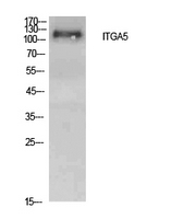
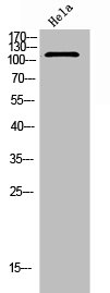
Figure 1. Western blot analysis of ITGA5 using anti-ITGA5 antibody (A01911). Electrophoresis was performed on a 5-20% SDS-PAGE gel at 70V (Stacking gel) / 90V (Resolving gel) for 2-3 hours. The sample well of each lane was loaded with 30 ug of sample under reducing conditions. Lane 1: human A549 whole cell lysates, Lane 2: human Hela whole cell lysates, Lane 3: human placenta tissue lysates, Lane 4: rat brain tissue lysates, Lane 5: rat PC-12 whole cell lysates, Lane 6: mouse brain tissue lysates. After electrophoresis, proteins were transferred to a nitrocellulose membrane at 150 mA for 50-90 minutes. Blocked the membrane with 5% non-fat milk/TBS for 1.5 hour at RT. The membrane was incubated with rabbit anti-ITGA5 antigen affinity purified polyclonal antibody (Catalog # A01911) at 0.5 microg/mL overnight at 4°C, then washed with TBS-0.1%Tween 3 times with 5 minutes each and probed with a goat anti-rabbit IgG-HRP secondary antibody at a dilution of 1:5000 for 1.5 hour at RT. The signal is developed using an Enhanced Chemiluminescent detection (ECL) kit (Catalog # EK1002) with Tanon 5200 system. A specific band was detected for ITGA5 at approximately 130 kDa. The expected band size for ITGA5 is at 115 kDa.
. ITGA5 was detected in a paraffin-embedded section of human placenta tissue. Heat mediated antigen retrieval was performed in EDTA buffer (pH 8.0, epitope retrieval solution). The tissue section was blocked with 10% goat serum. The tissue section was then incubated with 2 microg/ml rabbit anti-ITGA5 Antibody (A01911) overnight at 4°C. Peroxidase Conjugated Goat Anti-rabbit IgG was used as secondary antibody and incubated for 30 minutes at 37°C. The tissue section was developed using HRP Conjugated Rabbit IgG Super Vision Assay Kit (Catalog # SV0002) with DAB as the chromogen.")
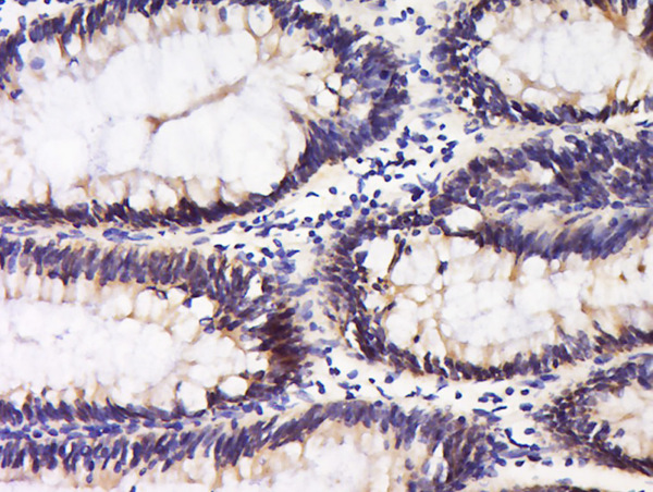
. ITGA5 was detected in a paraffin-embedded section of rat bladder tissue. Heat mediated antigen retrieval was performed in EDTA buffer (pH 8.0, epitope retrieval solution). The tissue section was blocked with 10% goat serum. The tissue section was then incubated with 2 microg/ml rabbit anti-ITGA5 Antibody (A01911) overnight at 4°C. Peroxidase Conjugated Goat Anti-rabbit IgG was used as secondary antibody and incubated for 30 minutes at 37°C. The tissue section was developed using HRP Conjugated Rabbit IgG Super Vision Assay Kit (Catalog # SV0002) with DAB as the chromogen.")
Figure 1. Western blot analysis of ITGA5 using anti-ITGA5 antibody (A01911). Electrophoresis was performed on a 5-20% SDS-PAGE gel at 70V (Stacking gel) / 90V (Resolving gel) for 2-3 hours. The sample well of each lane was loaded with 30 ug of sample under reducing conditions. Lane 1: human A549 whole cell lysates, Lane 2: human Hela whole cell lysates, Lane 3: human placenta tissue lysates, Lane 4: rat brain tissue lysates, Lane 5: rat PC-12 whole cell lysates, Lane 6: mouse brain tissue lysates. After electrophoresis, proteins were transferred to a nitrocellulose membrane at 150 mA for 50-90 minutes. Blocked the membrane with 5% non-fat milk/TBS for 1.5 hour at RT. The membrane was incubated with rabbit anti-ITGA5 antigen affinity purified polyclonal antibody (Catalog # A01911) at 0.5 microg/mL overnight at 4°C, then washed with TBS-0.1%Tween 3 times with 5 minutes each and probed with a goat anti-rabbit IgG-HRP secondary antibody at a dilution of 1:5000 for 1.5 hour at RT. The signal is developed using an Enhanced Chemiluminescent detection (ECL) kit (Catalog # EK1002) with Tanon 5200 system. A specific band was detected for ITGA5 at approximately 130 kDa. The expected band size for ITGA5 is at 115 kDa.
Anti-Integrin alpha 5/ITGA5 Antibody Picoband(r)
A01911-CARRIER-FREE
ApplicationsWestern Blot, ELISA, ImmunoHistoChemistry
Product group Antibodies
ReactivityHuman, Mouse, Rat
TargetITGA5
Overview
- SupplierBoster Bio
- Product NameAnti-Integrin alpha 5/ITGA5 Antibody Picoband(r)
- Delivery Days Customer9
- ApplicationsWestern Blot, ELISA, ImmunoHistoChemistry
- CertificationResearch Use Only
- ClonalityPolyclonal
- Concentration500 ug/ml
- Gene ID3678
- Target nameITGA5
- Target descriptionintegrin subunit alpha 5
- Target synonymsCD49e, FNRA, VLA-5, VLA5A, integrin alpha-5, CD49 antigen-like family member E, ITGA5/SLC9B1 fusion, fibronectin receptor subunit alpha, fibronectin receptor, alpha polypeptide, fibronectin receptor, alpha subunit, integrin alpha-F, integrin, alpha 5 (fibronectin receptor, alpha polypeptide), very late activation protein 5, alpha subunit
- HostRabbit
- IsotypeIgG
- Protein IDP08648
- Protein NameIntegrin alpha-5
- Scientific DescriptionBoster Bio Anti-Integrin alpha 5/ITGA5 Antibody Picoband® catalog # A01911. Tested in ELISA, IHC, WB applications. This antibody reacts with Human, Mouse, Rat. The brand Picoband indicates this is a premium antibody that guarantees superior quality, high affinity, and strong signals with minimal background in Western blot applications. Only our best-performing antibodies are designated as Picoband, ensuring unmatched performance.
- ReactivityHuman, Mouse, Rat
- Storage Instruction-20°C,2°C to 8°C
- UNSPSC12352203
Related products
Product group Antibodies
Anti-ITGA5 AntibodyA97455
ApplicationsWestern Blot, ELISA
ReactivityHuman, Mouse, Rat
- SizePrice
Product group Antibodies

ApplicationsFlow Cytometry, ImmunoFluorescence, ELISA, ImmunoHistoChemistry, ImmunoHistoChemistry Frozen, ImmunoHistoChemistry Paraffin, Neutralisation/Blocking
ReactivityHuman
TargetITGA5
- SizePrice
Product group Antibodies
Anti-ITGA5 Antibody144-06209
ApplicationsWestern Blot
ReactivityHuman, Mouse, Rat
TargetITGA5
- SizePrice
Product group Antibodies

CD49e AntibodyABX031763
ApplicationsFlow Cytometry, Western Blot, ELISA
- SizePrice

Product group Antibodies
Anti-ITGA5 AntibodyAMAB91447
ApplicationsWestern Blot, ImmunoHistoChemistry
ReactivityHuman
TargetITGA5
- SizePrice
Product group Antibodies
References
ApplicationsImmunoFluorescence, Western Blot, ELISA, ImmunoCytoChemistry, ImmunoHistoChemistry, ImmunoHistoChemistry Frozen, ImmunoHistoChemistry Paraffin
ReactivityBovine, Equine, Human, Mouse, Porcine, Rabbit, Rat, Sheep
TargetITGA5
- SizePrice
Product group Antibodies
ITGA5 AntibodyCSB-PA006198
ApplicationsWestern Blot, ELISA
ReactivityHuman, Mouse, Rat
TargetITGA5
- SizePrice
Product group Antibodies
Itga5 Polyclonal AntibodyCAC08565
ApplicationsImmunoFluorescence, Western Blot, ELISA, ImmunoHistoChemistry
ReactivityMouse
TargetITGA5
- SizePrice
Product group Antibodies

ApplicationsFlow Cytometry, ImmunoPrecipitation, ImmunoCytoChemistry, ImmunoHistoChemistry, ImmunoHistoChemistry Frozen
ReactivityHuman, Primate
TargetITGA5
- SizePrice