

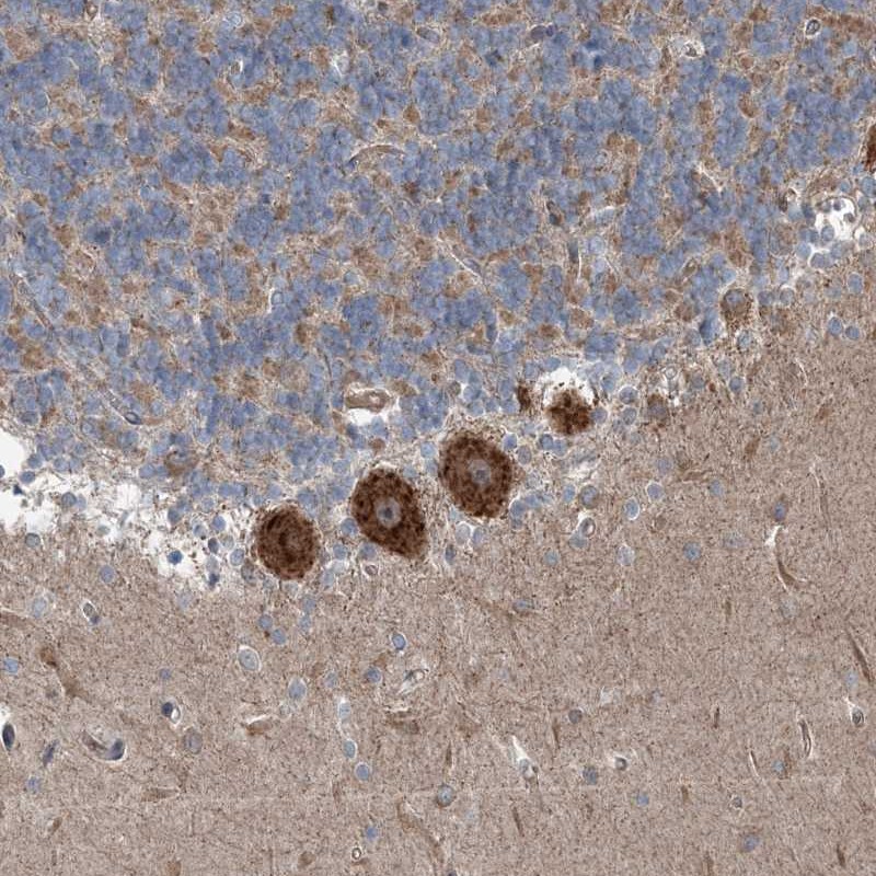
Immunohistochemical staining of human cerebellum shows strong cytoplasmic positivity in purkinje cells.
Immunohistochemical staining of human cerebellum shows strong cytoplasmic positivity in purkinje cells.
Anti-INTU Antibody
HPA036714
ApplicationsImmunoHistoChemistry
Product group Antibodies
ReactivityHuman
TargetINTU
Overview
- SupplierAtlas Antibodies
- Product NameAnti-INTU Antibody
- Delivery Days Customer4
- ApplicationsImmunoHistoChemistry
- CertificationResearch Use Only
- ClonalityPolyclonal
- ConjugateUnconjugated
- Gene ID27152
- Target nameINTU
- Target descriptioninturned planar cell polarity protein
- Target synonymsCPLANE4, INT, OFD17, PDZD6, PDZK6, SRTD20, protein inturned, PDZ domain containing 6, PDZ domain-containing protein 6, ciliogenesis and planar polarity effector 4, ciliogenesis and planar polarity effector complex subunit 4, inturned planar cell polarity effector homolog
- HostRabbit
- IsotypeIgG
- Protein IDQ9ULD6
- Protein NameProtein inturned
- Scientific DescriptionRecombinant Protein Epitope Signature Tag (PrEST) antigen sequence
- ReactivityHuman
- Storage Instruction-20°C,2°C to 8°C
- UNSPSC41116161
Datasheet
MSDS
Related products

Product group Antibodies
Anti-INTU Antibody Picoband(r)A11533-1-CARRIER-FREE
ApplicationsFlow Cytometry, ImmunoFluorescence, Western Blot, ELISA, ImmunoHistoChemistry
ReactivityHuman
TargetINTU
- SizePrice

Product group Antibodies
Anti-INTU AntibodyHPA036715
ApplicationsImmunoHistoChemistry
ReactivityHuman
TargetINTU
- SizePrice
![Lane 1: Marker [kDa] 250, 130, 95, 72, 55, 36, 28, 17, 10 | Lane 2: RT4 | Lane 3: U-251 MG | Lane 4: Human Plasma | Lane 5: Liver | Lane 6: Tonsil](https://atlasantibodies.s3.amazonaws.com/images/wb/hpa061467-wb-1.jpg)
Product group Antibodies
Anti-INTU AntibodyHPA061467
ApplicationsWestern Blot
ReactivityHuman
TargetINTU
- SizePrice
Product group Antibodies

INTU AntibodyLS-C680678
ApplicationsImmunoFluorescence, ELISA, ImmunoHistoChemistry, ImmunoHistoChemistry Paraffin
ReactivityHuman
TargetINTU
- SizePrice
Product group Antibodies
INTU AntibodyCSB-PA891544LA01HU
ApplicationsImmunoFluorescence, ELISA, ImmunoHistoChemistry
ReactivityHuman
TargetINTU
- SizePrice
Product group Antibodies
INTU AntibodyPACO60436
ApplicationsImmunoFluorescence, ELISA, ImmunoHistoChemistry
ReactivityHuman
TargetINTU
- SizePrice
Product group Antibodies
PDZD6 Polyclonal AntibodyBS-9041R
ApplicationsWestern Blot
ReactivityHuman, Mouse, Rabbit, Rat
TargetINTU
- SizePrice