

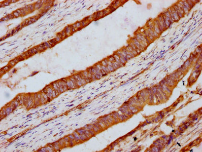
IHC image of CSB-PA891544LA01HU diluted at 1:300 and staining in paraffin-embedded human colon cancer performed on a Leica BondTM system. After dewaxing and hydration, antigen retrieval was mediated by high pressure in a citrate buffer (pH 6.0). Section was blocked with 10% normal goat serum 30min at RT. Then primary antibody (1% BSA) was incubated at 4°C overnight. The primary is detected by a biotinylated secondary antibody and visualized using an HRP conjugated SP system.
. Section was blocked with 10% normal goat serum 30min at RT. Then primary antibody (1% BSA) was incubated at 4°C overnight. The primary is detected by a biotinylated secondary antibody and visualized using an HRP conjugated SP system.")
.")
IHC image of CSB-PA891544LA01HU diluted at 1:300 and staining in paraffin-embedded human colon cancer performed on a Leica BondTM system. After dewaxing and hydration, antigen retrieval was mediated by high pressure in a citrate buffer (pH 6.0). Section was blocked with 10% normal goat serum 30min at RT. Then primary antibody (1% BSA) was incubated at 4°C overnight. The primary is detected by a biotinylated secondary antibody and visualized using an HRP conjugated SP system.
INTU Antibody
CSB-PA891544LA01HU
ApplicationsImmunoFluorescence, ELISA, ImmunoHistoChemistry
Product group Antibodies
ReactivityHuman
TargetINTU
Overview
- SupplierCusabio
- Product NameINTU Antibody
- Delivery Days Customer20
- ApplicationsImmunoFluorescence, ELISA, ImmunoHistoChemistry
- CertificationResearch Use Only
- ClonalityPolyclonal
- ConjugateUnconjugated
- Gene ID27152
- Target nameINTU
- Target descriptioninturned planar cell polarity protein
- Target synonymsCPLANE4, INT, OFD17, PDZD6, PDZK6, SRTD20, protein inturned, PDZ domain containing 6, PDZ domain-containing protein 6, ciliogenesis and planar polarity effector 4, ciliogenesis and planar polarity effector complex subunit 4, inturned planar cell polarity effector homolog
- HostRabbit
- IsotypeIgG
- Protein IDQ9ULD6
- Protein NameProtein inturned
- Scientific DescriptionPlays a key role in ciliogenesis and embryonic development. Regulator of cilia formation by controlling the organization of the apical actin cytoskeleton and the positioning of the basal bodies at the apical cell surface, which in turn is essential for the normal orientation of elongating ciliary microtubules. Plays a key role in definition of cell polarity via its role in ciliogenesis but not via conversion extension. Has an indirect effect on hedgehog signaling (By similarity). Proposed to function as core component of the CPLANE (ciliogenesis and planar polarity effectors) complex involved in the recruitment of peripheral IFT-A proteins to basal bodies (PubMed:27158779).
- ReactivityHuman
- Storage Instruction-20°C or -80°C
- UNSPSC41116161
Related products

Product group Antibodies
Anti-INTU Antibody Picoband(r)A11533-1-CARRIER-FREE
ApplicationsFlow Cytometry, ImmunoFluorescence, Western Blot, ELISA, ImmunoHistoChemistry
ReactivityHuman
TargetINTU
- SizePrice
Product group Antibodies
Anti-INTU AntibodyHPA036714
ApplicationsImmunoHistoChemistry
ReactivityHuman
TargetINTU
- SizePrice
Product group Antibodies

INTU AntibodyLS-C680678
ApplicationsImmunoFluorescence, ELISA, ImmunoHistoChemistry, ImmunoHistoChemistry Paraffin
ReactivityHuman
TargetINTU
- SizePrice
Product group Antibodies
INTU AntibodyPACO60436
ApplicationsImmunoFluorescence, ELISA, ImmunoHistoChemistry
ReactivityHuman
TargetINTU
- SizePrice
Product group Antibodies
PDZD6 Polyclonal AntibodyBS-9041R
ApplicationsWestern Blot
ReactivityHuman, Mouse, Rabbit, Rat
TargetINTU
- SizePrice
Product group Antibodies
INTU antibodyGTX120239
ApplicationsImmunoFluorescence, Western Blot, ImmunoCytoChemistry
ReactivityHuman, Mouse
TargetINTU
- SizePrice