Bio-Connect
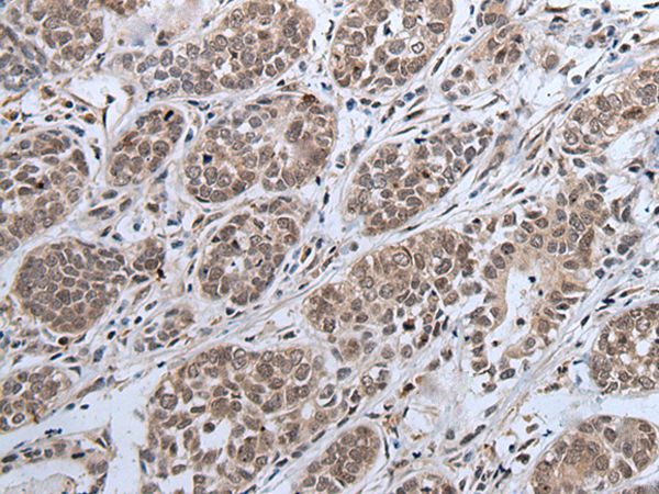
Immunohistochemical staining of human tonsil shows strong nuclear positivity in reaction center cells.

Anti-IWS1 Antibody

HPA035719
Atlas Antibodies
ApplicationsImmunoHistoChemistry
Product group Antibodies
TargetIWS1
Sign in to order and to see your custom pricing.
Large volume orders?
Order with a bulk request

Overview

  • Supplier
    Atlas Antibodies
  • Product Name
    Anti-IWS1
  • Delivery Days Customer
    4
  • Applications
    ImmunoHistoChemistry
  • Certification
    Research Use Only
  • Clonality
    Polyclonal
  • Conjugate
    Unconjugated
  • Gene ID55677
  • Target name
    IWS1
  • Target description
    interacts with SUPT6H, CTD assembly factor 1
  • Target synonyms
    interacts with Spt6; IWS1 homolog; IWS1, SUPT6H interacting protein; IWS1-like protein; protein IWS1 homolog
  • Host
    Rabbit
  • Isotype
    IgG
  • Protein IDQ96ST2
  • Protein Name
    Protein IWS1 homolog
  • Scientific Description
    Recombinant Protein Epitope Signature Tag (PrEST) antigen sequence
  • Storage Instruction
    -20°C,2°C to 8°C
  • UNSPSC
    12352203

Related products

Western blot All lanes: IWS1 antibody at 3.84microg/ml Lane 1: 293T whole cell lysate Lane 2: Jurkat whole cell lysate Secondary Goat polyclonal to rabbit IgG at 1/10000 dilution Predicted band size: 92, 57, 70 kDa Observed band size: 70 kDa
Product group Antibodies
Cusabio
IWS1 AntibodyCSB-PA822304ESR1HU
ApplicationsWestern Blot, ELISA, ImmunoHistoChemistry
TargetIWS1
  • SizePrice
Immunohistochemistry analysis in human placenta and pancreas tissues using Anti-IWS1 antibody. Corresponding IWS1 RNA-seq data are presented for the same tissues.
Product group Antibodies
Atlas Antibodies
ApplicationsImmunoCytoChemistry, ImmunoHistoChemistry
TargetIWS1
  • SizePrice
Atlas Antibodies
Protein IDQ96ST2
  • SizePrice
Product group Antibodies
Bethyl Laboratories
ApplicationsImmunoPrecipitation, Western Blot
TargetIWS1
  • SizePrice
Atlas Antibodies
Protein IDQ96ST2
  • SizePrice
Figure 1. Western blot analysis of IWS1 using anti-IWS1 antibody (A05958-1). Electrophoresis was performed on a 5-20% SDS-PAGE gel at 70V (Stacking gel) / 90V (Resolving gel) for 2-3 hours. The sample well of each lane was loaded with 30 ug of sample under reducing conditions. Lane 1: human Hela whole cell lysates, Lane 2: human HEK293 whole cell lysates, Lane 3: human Jurkat whole cell lysates, Lane 4: human HepG2 whole cell lysates, Lane 5: rat C6 whole cell lysates. After electrophoresis, proteins were transferred to a nitrocellulose membrane at 150 mA for 50-90 minutes. Blocked the membrane with 5% non-fat milk/TBS for 1.5 hour at RT. The membrane was incubated with rabbit anti-IWS1 antigen affinity purified polyclonal antibody (Catalog # A05958-1) at 0.25 microg/mL overnight at 4°C, then washed with TBS-0.1%Tween 3 times with 5 minutes each and probed with a goat anti-rabbit IgG-HRP secondary antibody at a dilution of 1:5000 for 1.5 hour at RT. The signal is developed using an Enhanced Chemiluminescent detection (ECL) kit (Catalog # EK1002) with Tanon 5200 system. A specific band was detected for IWS1 at approximately 140 kDa. The expected band size for IWS1 is at 140 kDa.
Product group Antibodies
Boster Bio
Anti-IWS1 Antibody Picoband(r)A05958-1-CARRIER-FREE
ApplicationsFlow Cytometry, Western Blot, ELISA
TargetIWS1
  • SizePrice
Product group Antibodies
LifeSpan BioSciences
IWS1 AntibodyLS-C349095
ApplicationsImmunoFluorescence, Western Blot, ImmunoHistoChemistry
TargetIWS1
  • SizePrice
Product group Antibodies
Applied Biological Materials
Anti-IWS1Y058896
ApplicationsWestern Blot, ELISA, ImmunoHistoChemistry
  • SizePrice
Anti-IWS1 Antibody
Product group Antibodies
Antibodies.com
ApplicationsImmunoHistoChemistry
  • SizePrice