


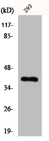
Figure 1. Western blot analysis of JAB1 using anti-JAB1 antibody (PB9490). Electrophoresis was performed on a 5-20% SDS-PAGE gel at 70V (Stacking gel) / 90V (Resolving gel) for 2-3 hours. The sample well of each lane was loaded with 30 ug of sample under reducing conditions. Lane 1: human Raji whole cell lysates, Lane 2: human K562 whole cell lysates, Lane 3: human PC-3 whole cell lysates, Lane 4: human Hela whole cell lysates, Lane 5: human A549 whole cell lysates, Lane 6: human HEK293 whole cell lysates, Lane 7: rat brain tissue lysates, Lane 8: rat heart tissue lysates, Lane 9: mouse brain tissue lysates, Lane 10: mouse heart tissue lysates, Lane 11: mouse SP2/0 whole cell lysates. After electrophoresis, proteins were transferred to a nitrocellulose membrane at 150 mA for 50-90 minutes. Blocked the membrane with 5% non-fat milk/TBS for 1.5 hour at RT. The membrane was incubated with rabbit anti-JAB1 antigen affinity purified polyclonal antibody (Catalog # PB9490) at 0.5 microg/mL overnight at 4°C, then washed with TBS-0.1%Tween 3 times with 5 minutes each and probed with a goat anti-rabbit IgG-HRP secondary antibody at a dilution of 1:5000 for 1.5 hour at RT. The signal is developed using an Enhanced Chemiluminescent detection (ECL) kit (Catalog # EK1002) with Tanon 5200 system. A specific band was detected for JAB1 at approximately 38 kDa. The expected band size for JAB1 is at 38 kDa.
. JAB1 was detected in a paraffin-embedded section of mouse testis tissue. Heat mediated antigen retrieval was performed in EDTA buffer (pH 8.0, epitope retrieval solution). The tissue section was blocked with 10% goat serum. The tissue section was then incubated with 1 microg/ml rabbit anti-JAB1 Antibody (PB9490) overnight at 4°C. Biotinylated goat anti-rabbit IgG was used as secondary antibody and incubated for 30 minutes at 37°C. The tissue section was developed using Strepavidin-Biotin-Complex (SABC) (Catalog # SA1022) with DAB as the chromogen.")
. JAB1 was detected in a paraffin-embedded section of rat testis tissue. Heat mediated antigen retrieval was performed in EDTA buffer (pH 8.0, epitope retrieval solution). The tissue section was blocked with 10% goat serum. The tissue section was then incubated with 1 microg/ml rabbit anti-JAB1 Antibody (PB9490) overnight at 4°C. Biotinylated goat anti-rabbit IgG was used as secondary antibody and incubated for 30 minutes at 37°C. The tissue section was developed using Strepavidin-Biotin-Complex (SABC) (Catalog # SA1022) with DAB as the chromogen.")
. JAB1 was detected in a paraffin-embedded section of human mammary cancer tissue. Heat mediated antigen retrieval was performed in EDTA buffer (pH 8.0, epitope retrieval solution). The tissue section was blocked with 10% goat serum. The tissue section was then incubated with 1 microg/ml rabbit anti-JAB1 Antibody (PB9490) overnight at 4°C. Biotinylated goat anti-rabbit IgG was used as secondary antibody and incubated for 30 minutes at 37°C. The tissue section was developed using Strepavidin-Biotin-Complex (SABC) (Catalog # SA1022) with DAB as the chromogen.")
. Overlay histogram showing U20S cells stained with PB9490 (Blue line). To facilitate intracellular staining, cells were fixed with 4% paraformaldehyde and permeabilized with permeabilization buffer. The cells were blocked with 10% normal goat serum. And then incubated with rabbit anti-JAB1 Antibody (PB9490,1microg/1x106 cells) for 30 min at 20°C. DyLight®488 conjugated goat anti-rabbit IgG (BA1127, 5-10microg/1x106 cells) was used as secondary antibody for 30 minutes at 20°C. Isotype control antibody (Green line) was rabbit IgG (1microg/1x106) used under the same conditions. Unlabelled sample without incubation with primary antibody and secondary antibody (Red line) was used as a blank control.")
. Overlay histogram showing U937 cells stained with PB9490 (Blue line). To facilitate intracellular staining, cells were fixed with 4% paraformaldehyde and permeabilized with permeabilization buffer. The cells were blocked with 10% normal goat serum. And then incubated with rabbit anti-JAB1 Antibody (PB9490,1microg/1x106 cells) for 30 min at 20°C. DyLight®488 conjugated goat anti-rabbit IgG (BA1127, 5-10microg/1x106 cells) was used as secondary antibody for 30 minutes at 20°C. Isotype control antibody (Green line) was rabbit IgG (1microg/1x106) used under the same conditions. Unlabelled sample without incubation with primary antibody and secondary antibody (Red line) was used as a blank control.")
. JAB1 was detected in immunocytochemical section of A431 cell. Enzyme antigen retrieval was performed using IHC enzyme antigen retrieval reagent (AR0022) for 15 mins. The cells were blocked with 10% goat serum. And then incubated with 1microg/ml rabbit anti-JAB1 Antibody (PB9490) overnight at 4°C. Biotinylated goat anti-rabbit IgG was used as secondary antibody and incubated for 30 minutes at 37°C. The section was developed using Strepavidin-Biotin-Complex (SABC)(Catalog # SA1022) with DAB as the chromogen.")
Figure 1. Western blot analysis of JAB1 using anti-JAB1 antibody (PB9490). Electrophoresis was performed on a 5-20% SDS-PAGE gel at 70V (Stacking gel) / 90V (Resolving gel) for 2-3 hours. The sample well of each lane was loaded with 30 ug of sample under reducing conditions. Lane 1: human Raji whole cell lysates, Lane 2: human K562 whole cell lysates, Lane 3: human PC-3 whole cell lysates, Lane 4: human Hela whole cell lysates, Lane 5: human A549 whole cell lysates, Lane 6: human HEK293 whole cell lysates, Lane 7: rat brain tissue lysates, Lane 8: rat heart tissue lysates, Lane 9: mouse brain tissue lysates, Lane 10: mouse heart tissue lysates, Lane 11: mouse SP2/0 whole cell lysates. After electrophoresis, proteins were transferred to a nitrocellulose membrane at 150 mA for 50-90 minutes. Blocked the membrane with 5% non-fat milk/TBS for 1.5 hour at RT. The membrane was incubated with rabbit anti-JAB1 antigen affinity purified polyclonal antibody (Catalog # PB9490) at 0.5 microg/mL overnight at 4°C, then washed with TBS-0.1%Tween 3 times with 5 minutes each and probed with a goat anti-rabbit IgG-HRP secondary antibody at a dilution of 1:5000 for 1.5 hour at RT. The signal is developed using an Enhanced Chemiluminescent detection (ECL) kit (Catalog # EK1002) with Tanon 5200 system. A specific band was detected for JAB1 at approximately 38 kDa. The expected band size for JAB1 is at 38 kDa.
Anti-JAB1/COPS5 Antibody Picoband(r)
PB9490-CARRIER-FREE
ApplicationsFlow Cytometry, ImmunoFluorescence, ImmunoPrecipitation, Western Blot, ImmunoCytoChemistry, ImmunoHistoChemistry, ImmunoHistoChemistry Frozen
Product group Antibodies
ReactivityBovine, Canine, Chicken, Equine, Human, Mouse, Rabbit, Rat
TargetCOPS5
Overview
- SupplierBoster Bio
- Product NameAnti-JAB1/COPS5 Antibody Picoband(r)
- Delivery Days Customer9
- Application Supplier NoteTested Species: In-house tested species with positive results. By Heat: Boiling the paraffin sections in 10mM citrate buffer, pH6.0, for 20mins is required for the staining of formalin/paraffin sections. Other applications have not been tested. Optimal dilutions should be determined by end users.
- ApplicationsFlow Cytometry, ImmunoFluorescence, ImmunoPrecipitation, Western Blot, ImmunoCytoChemistry, ImmunoHistoChemistry, ImmunoHistoChemistry Frozen
- CertificationResearch Use Only
- ClonalityPolyclonal
- Concentration500 ug/ml
- Gene ID10987
- Target nameCOPS5
- Target descriptionCOP9 signalosome subunit 5
- Target synonymsCSN5, JAB1, MOV-34, SGN5, COP9 signalosome complex subunit 5, 38 kDa Mov34 homolog, COP9 constitutive photomorphogenic homolog subunit 5, jun activation domain-binding protein 1, signalosome subunit 5, testis secretory sperm-binding protein Li 231m
- HostRabbit
- IsotypeIgG
- Protein IDQ92905
- Protein NameCOP9 signalosome complex subunit 5
- Scientific DescriptionBoster Bio Anti-JAB1/COPS5 Antibody Picoband® catalog # PB9490. Tested in Flow Cytometry, IP, IF, IHC, IHC-F, ICC, WB applications. This antibody reacts with Human, Mouse, Rat. The brand Picoband indicates this is a premium antibody that guarantees superior quality, high affinity, and strong signals with minimal background in Western blot applications. Only our best-performing antibodies are designated as Picoband, ensuring unmatched performance.
- ReactivityBovine, Canine, Chicken, Equine, Human, Mouse, Rabbit, Rat
- Storage Instruction-20°C,2°C to 8°C
- UNSPSC12352203
Related products
Product group Antibodies
Anti-COPS5 AntibodyA99133
ApplicationsWestern Blot, ELISA
ReactivityHuman, Mouse
- SizePrice
Product group Antibodies
Anti-COPS5 Antibody144-01766
ApplicationsWestern Blot, ImmunoHistoChemistry
ReactivityHuman, Mouse, Rat
TargetCOPS5
- SizePrice
Product group Antibodies
Jab1/COPS5 Recombinant Antibody, Biotin ConjugatedBSM-61721R-BIOTIN
ApplicationsImmunoPrecipitation, Western Blot, ImmunoHistoChemistry, ImmunoHistoChemistry Frozen, ImmunoHistoChemistry Paraffin
ReactivityHuman, Mouse, Rat
TargetCOPS5
- SizePrice
Product group Antibodies
COPS5 AntibodyCSB-PA003075
ApplicationsWestern Blot, ELISA, ImmunoHistoChemistry
ReactivityHuman, Mouse
TargetCOPS5
- SizePrice
Product group Antibodies

COPS5 / JAB1 AntibodyLS-C400839
ApplicationsWestern Blot, ELISA, ImmunoHistoChemistry
ReactivityHuman, Mouse
TargetCOPS5
- SizePrice

Product group Antibodies
Anti-COPS5 AntibodyHPA004845
ApplicationsWestern Blot, ImmunoCytoChemistry, ImmunoHistoChemistry
ReactivityHuman, Mouse, Rat
TargetCOPS5
- SizePrice
![Jab1 antibody [N1C1] detects Jab1 protein on zebrafish by whole mount immunohistochemical analysis. Sample: 2 days-post-fertilization zebrafish embryo. Jab1 antibody [N1C1] (GTX113927) dilution: 1:100.](https://www.genetex.com/upload/website/prouct_img/normal/GTX113927/GTX113927_40150_20150427_IHC-Wm_Z_22111423_536.webp)
Product group Antibodies
Jab1 antibody [N1C1]GTX113927
ApplicationsImmunoFluorescence, Western Blot, ImmunoCytoChemistry, ImmunoHistoChemistry, ImmunoHistoChemistry Paraffin
ReactivityHuman, Mouse, Zebra Fish
TargetCOPS5
- SizePrice