

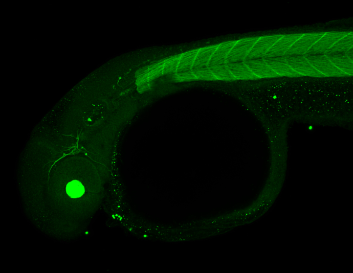
Jab1 antibody [N1C1] detects Jab1 protein on zebrafish by whole mount immunohistochemical analysis. Sample: 2 days-post-fertilization zebrafish embryo. Jab1 antibody [N1C1] (GTX113927) dilution: 1:100.
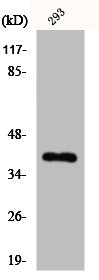
 A: Zebrafish eye 10% SDS PAGE GTX113927 diluted at 1:1000")
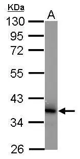
 A: Hela 10% SDS PAGE GTX113927 diluted at 1:1000")
![JAB1 antibody [N1C1] detects JAB1 protein at nucleus by immunofluorescent analysis. Sample: HeLa cells were fixed in 4% paraformaldehyde at RT for 15 min. Green: JAB1 protein stained by JAB1 antibody [N1C1] (GTX113927) diluted at 1:200. Red: FIS1, a mitochondria marker, stained by FIS1 antibody [GT9810] (GTX631209) diluted at 1:20000. Blue: Hoechst 33342 staining.](https://www.genetex.com/upload/website/prouct_img/normal/GTX113927/GTX113927_40150_20150410_IFA_w_23060501_483.webp "JAB1 antibody [N1C1] detects JAB1 protein at nucleus by immunofluorescent analysis. Sample: HeLa cells were fixed in 4% paraformaldehyde at RT for 15 min. Green: JAB1 protein stained by JAB1 antibody [N1C1] (GTX113927) diluted at 1:200. Red: FIS1, a mitochondria marker, stained by FIS1 antibody [GT9810] (GTX631209) diluted at 1:20000. Blue: Hoechst 33342 staining.")
![JAB1 antibody [N1C1] detects JAB1 protein at nucleus on mouse middle brain by immunohistochemical analysis. Sample: Paraffin-embedded mouse middle brain. JAB1 antibody [N1C1] (GTX113927) dilution: 1:500.
Antigen Retrieval: Trilogy? (EDTA based, pH 8.0) buffer, 15min](https://www.genetex.com/upload/website/prouct_img/normal/GTX113927/GTX113927_40150_IHC_M_w_23060501_401.webp "JAB1 antibody [N1C1] detects JAB1 protein at nucleus on mouse middle brain by immunohistochemical analysis. Sample: Paraffin-embedded mouse middle brain. JAB1 antibody [N1C1] (GTX113927) dilution: 1:500.
Antigen Retrieval: Trilogy? (EDTA based, pH 8.0) buffer, 15min")
 A: Mouse brain 10% SDS PAGE GTX113927 diluted at 1:1000")
Jab1 antibody [N1C1] detects Jab1 protein on zebrafish by whole mount immunohistochemical analysis. Sample: 2 days-post-fertilization zebrafish embryo. Jab1 antibody [N1C1] (GTX113927) dilution: 1:100.
Jab1 antibody [N1C1]
GTX113927
ApplicationsImmunoFluorescence, Western Blot, ImmunoCytoChemistry, ImmunoHistoChemistry, ImmunoHistoChemistry Paraffin
Product group Antibodies
ReactivityHuman, Mouse, Zebra Fish
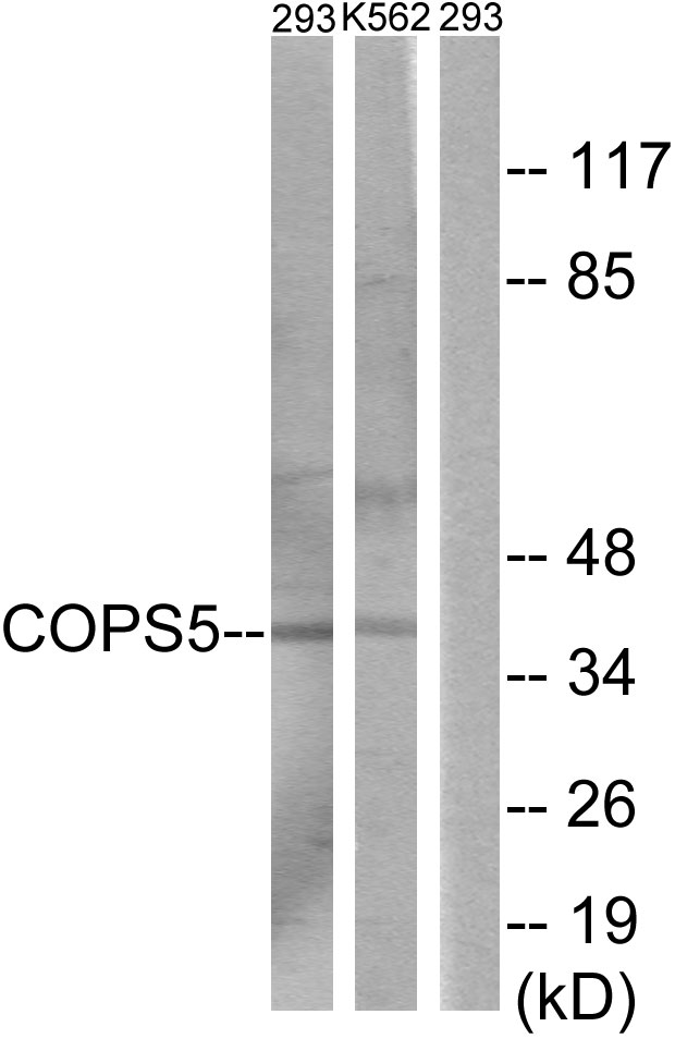
TargetCOPS5
Overview
- SupplierGeneTex
- Product NameJab1 antibody [N1C1]
- Delivery Days Customer9
- Application Supplier NoteWB: 1:500-1:3000. ICC/IF: 1:100-1:1000. IHC-P: 1:100-1:1000. *Optimal dilutions/concentrations should be determined by the researcher.Not tested in other applications.
- ApplicationsImmunoFluorescence, Western Blot, ImmunoCytoChemistry, ImmunoHistoChemistry, ImmunoHistoChemistry Paraffin
- CertificationResearch Use Only
- ClonalityPolyclonal
- Concentration0.35 mg/ml
- ConjugateUnconjugated
- Gene ID10987
- Target nameCOPS5
- Target descriptionCOP9 signalosome subunit 5
- Target synonymsCSN5, JAB1, MOV-34, SGN5, COP9 signalosome complex subunit 5, 38 kDa Mov34 homolog, COP9 constitutive photomorphogenic homolog subunit 5, jun activation domain-binding protein 1, signalosome subunit 5, testis secretory sperm-binding protein Li 231m
- HostRabbit
- IsotypeIgG
- Protein IDQ92905
- Protein NameCOP9 signalosome complex subunit 5
- Scientific DescriptionThe protein encoded by this gene is one of the eight subunits of COP9 signalosome, a highly conserved protein complex that functions as an important regulator in multiple signaling pathways. The structure and function of COP9 signalosome is similar to that of the 19S regulatory particle of 26S proteasome. COP9 signalosome has been shown to interact with SCF-type E3 ubiquitin ligases and act as a positive regulator of E3 ubiquitin ligases. This protein is reported to be involved in the degradation of cyclin-dependent kinase inhibitor CDKN1B/p27Kip1. It is also known to be an coactivator that increases the specificity of JUN/AP1 transcription factors. [provided by RefSeq]
- ReactivityHuman, Mouse, Zebra Fish
- Storage Instruction-20°C or -80°C,2°C to 8°C
- UNSPSC41116161
Datasheet
Related products
Product group Antibodies
Anti-COPS5 AntibodyA99133
ApplicationsWestern Blot, ELISA
ReactivityHuman, Mouse
- SizePrice
Product group Antibodies
Anti-COPS5 Antibody144-01766
ApplicationsWestern Blot, ImmunoHistoChemistry
ReactivityHuman, Mouse, Rat
TargetCOPS5
- SizePrice
Product group Antibodies
Jab1/COPS5 Recombinant Antibody, Biotin ConjugatedBSM-61721R-BIOTIN
ApplicationsImmunoPrecipitation, Western Blot, ImmunoHistoChemistry, ImmunoHistoChemistry Frozen, ImmunoHistoChemistry Paraffin
ReactivityHuman, Mouse, Rat
TargetCOPS5
- SizePrice
Product group Antibodies
COPS5 AntibodyCSB-PA003075
ApplicationsWestern Blot, ELISA, ImmunoHistoChemistry
ReactivityHuman, Mouse
TargetCOPS5
- SizePrice
Product group Antibodies

COPS5 / JAB1 AntibodyLS-C400839
ApplicationsWestern Blot, ELISA, ImmunoHistoChemistry
ReactivityHuman, Mouse
TargetCOPS5
- SizePrice

Product group Antibodies
Anti-COPS5 AntibodyHPA004845
ApplicationsWestern Blot, ImmunoCytoChemistry, ImmunoHistoChemistry
ReactivityHuman, Mouse, Rat
TargetCOPS5
- SizePrice
Product group Antibodies
Jab1 antibodyGTX12185
ApplicationsImmunoPrecipitation, Western Blot, ImmunoHistoChemistry, ImmunoHistoChemistry Paraffin
ReactivityHuman, Mouse, Rat
TargetCOPS5
- SizePrice
Product group Antibodies
Jab1 antibody [2A10.8]GTX70203
ApplicationsImmunoFluorescence, ImmunoPrecipitation, Western Blot, ImmunoCytoChemistry, ImmunoHistoChemistry
ReactivityDrosophila, Human, Mouse, Rat
TargetCOPS5
- SizePrice
![Jab1 antibody [6C3.38]](https://www.genetex.com/upload/website/prouct_img/normal/GTX70205/Jab1-antibody-6C3.38-GTX70205-IHC-1_w_23061221_598.webp)
Product group Antibodies
Jab1 antibody [6C3.38]GTX70205
ApplicationsImmunoPrecipitation, Western Blot, ImmunoHistoChemistry
ReactivityHuman, Mouse
TargetCOPS5
- SizePrice
Product group Antibodies
Jab1 antibody [8H8.5]GTX70207
ApplicationsImmunoPrecipitation, Western Blot
ReactivityDrosophila, Human, Mouse
TargetCOPS5
- SizePrice