


Immunofluorescent staining of human cell line A-431 shows localization to vesicles.
Immunofluorescent staining of human cell line A-431 shows localization to vesicles.
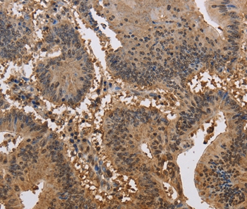
Anti-JAG2 Antibody
HPA050567
ApplicationsImmunoCytoChemistry
Product group Antibodies
ReactivityHuman
TargetJAG2
Overview
- SupplierAtlas Antibodies
- Product NameAnti-JAG2 Antibody
- Delivery Days Customer4
- ApplicationsImmunoCytoChemistry
- CertificationResearch Use Only
- ClonalityPolyclonal
- ConjugateUnconjugated
- Gene ID3714
- Target nameJAG2
- Target descriptionjagged canonical Notch ligand 2
- Target synonymsHJ2, LGMDR27, SER2, protein jagged-2, jagged 2
- HostRabbit
- IsotypeIgG
- Protein IDQ9Y219
- Protein NameProtein jagged-2
- Scientific DescriptionRecombinant Protein Epitope Signature Tag (PrEST) antigen sequence
- ReactivityHuman
- Storage Instruction-20°C,2°C to 8°C
- UNSPSC41116161
Datasheet
MSDS
Related products
Product group Antibodies
Anti-JAG2 AntibodyA46940
ApplicationsImmunoHistoChemistry
ReactivityHuman
- SizePrice
Product group Antibodies
Anti-JAG2 Antibody144-63149
ApplicationsImmunoFluorescence, Western Blot
ReactivityHuman, Mouse, Rat
TargetJAG2
- SizePrice

Product group Antibodies
Anti-Jagged 2/JAG2 Antibody Picoband(r)A01428-1-CARRIER-FREE
ApplicationsWestern Blot, ELISA
ReactivityHuman
TargetJAG2
- SizePrice
Product group Antibodies
Jagged 2 Polyclonal AntibodyBS-4244R
ApplicationsImmunoFluorescence, Western Blot, ELISA, ImmunoHistoChemistry, ImmunoHistoChemistry Frozen, ImmunoHistoChemistry Paraffin
ReactivityBovine, Canine, Human, Mouse, Porcine, Rat
TargetJAG2
- SizePrice
Product group Antibodies
ApplicationsImmunoPrecipitation, Western Blot, ImmunoCytoChemistry, ImmunoHistoChemistry
TargetJAG2
- SizePrice

Product group Antibodies
JAG2 AntibodyCSB-PA049990
ApplicationsELISA, ImmunoHistoChemistry
ReactivityHuman
TargetJAG2
- SizePrice
Product group Antibodies

JAG2 / Jagged-2 AntibodyLS-C403263
ApplicationsELISA, ImmunoHistoChemistry
ReactivityHuman
TargetJAG2
- SizePrice

Product group Antibodies
Anti-JAG2 AntibodyHPA030636
ApplicationsImmunoHistoChemistry
ReactivityHuman
TargetJAG2
- SizePrice