


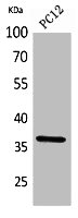
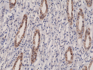
Figure 1. Western blot analysis of JAM-A/F11R using anti-JAM-A/F11R antibody (A02068-2). Electrophoresis was performed on a 5-20% SDS-PAGE gel at 70V (Stacking gel) / 90V (Resolving gel) for 2-3 hours. The sample well of each lane was loaded with 50ug of sample under reducing conditions. Lane 1: human placenta tissue lysates, Lane 2: human CACO-2 whole cell lysates, Lane 3: human MDA-MB-453 whole cell lysates, Lane 4: human HEPG2 whole cell lysates. After Electrophoresis, proteins were transferred to a Nitrocellulose membrane at 150mA for 50-90 minutes. Blocked the membrane with 5% Non-fat Milk/ TBS for 1.5 hour at RT. The membrane was incubated with rabbit anti-JAM-A/F11R antigen affinity purified polyclonal antibody (Catalog # A02068-2) at 0.5 microg/mL overnight at 4°C, then washed with TBS-0.1%Tween 3 times with 5 minutes each and probed with a goat anti-rabbit IgG-HRP secondary antibody at a dilution of 1:5000 for 1.5 hour at RT. The signal is developed using an Enhanced Chemiluminescent detection (ECL) kit (Catalog # EK1002) with Tanon 5200 system. A specific band was detected for JAM-A/F11R at approximately 41KD. The expected band size for JAM-A/F11R is at 41KD.
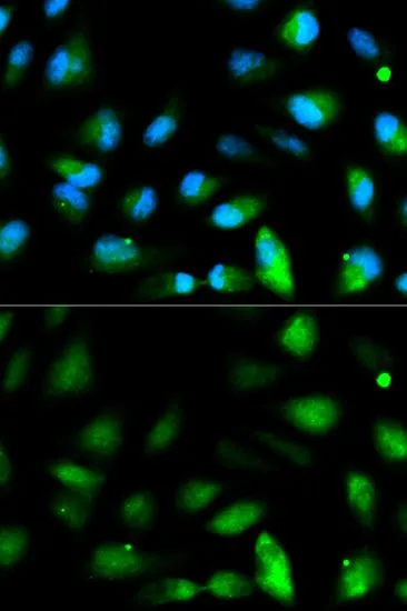
. JAM-A/F11R was detected in immunocytochemical section of MCF7 cells. Enzyme antigen retrieval was performed using IHC enzyme antigen retrieval reagent (AR0022) for 15 mins. The cells were blocked with 10% goat serum. And then incubated with 5microg/mL rabbit anti-JAM-A/F11R Antibody (A02068-2) overnight at 4°C. DyLight®594 Conjugated Goat Anti-Rabbit IgG (BA1142) was used as secondary antibody at 1:100 dilution and incubated for 30 minutes at 37°C. The section was counterstained with DAPI. Visualize using a fluorescence microscope and filter sets appropriate for the label used.")
. Overlay histogram showing HEPG2 cells stained with A02068-2 (Blue line). The cells were fixed with 4% paraformaldehyde and blocked with 10% normal goat serum. And then incubated with rabbit anti-JAM-A/F11R Antibody (A02068-2, 1microg/1x106 cells) for 30 min at 20°C. DyLight®488 conjugated goat anti-rabbit IgG (BA1127, 5-10microg/1x106 cells) was used as secondary antibody for 30 minutes at 20°C. Isotype control antibody (Green line) was rabbit IgG (1microg/1x106) used under the same conditions. Unlabelled sample without incubation with primary antibody and secondary antibody (Red line) was used as a blank control.")
Figure 1. Western blot analysis of JAM-A/F11R using anti-JAM-A/F11R antibody (A02068-2). Electrophoresis was performed on a 5-20% SDS-PAGE gel at 70V (Stacking gel) / 90V (Resolving gel) for 2-3 hours. The sample well of each lane was loaded with 50ug of sample under reducing conditions. Lane 1: human placenta tissue lysates, Lane 2: human CACO-2 whole cell lysates, Lane 3: human MDA-MB-453 whole cell lysates, Lane 4: human HEPG2 whole cell lysates. After Electrophoresis, proteins were transferred to a Nitrocellulose membrane at 150mA for 50-90 minutes. Blocked the membrane with 5% Non-fat Milk/ TBS for 1.5 hour at RT. The membrane was incubated with rabbit anti-JAM-A/F11R antigen affinity purified polyclonal antibody (Catalog # A02068-2) at 0.5 microg/mL overnight at 4°C, then washed with TBS-0.1%Tween 3 times with 5 minutes each and probed with a goat anti-rabbit IgG-HRP secondary antibody at a dilution of 1:5000 for 1.5 hour at RT. The signal is developed using an Enhanced Chemiluminescent detection (ECL) kit (Catalog # EK1002) with Tanon 5200 system. A specific band was detected for JAM-A/F11R at approximately 41KD. The expected band size for JAM-A/F11R is at 41KD.
Anti-JAM-A/F11R Antibody Picoband(r)
A02068-2-CARRIER-FREE
ApplicationsFlow Cytometry, ImmunoFluorescence, Western Blot, ImmunoCytoChemistry
Product group Antibodies
ReactivityHuman
TargetF11R
Overview
- SupplierBoster Bio
- Product NameAnti-JAM-A/F11R Antibody Picoband(r)
- Delivery Days Customer9
- ApplicationsFlow Cytometry, ImmunoFluorescence, Western Blot, ImmunoCytoChemistry
- CertificationResearch Use Only
- ClonalityPolyclonal
- Concentration500 ug/ml
- Gene ID50848
- Target nameF11R
- Target descriptionF11 receptor
- Target synonymsCD321, JAM, JAM1, JAMA, JCAM, KAT, PAM-1, junctional adhesion molecule A, junctional adhesion molecule 1, platelet F11 receptor, platelet adhesion molecule 1
- HostRabbit
- IsotypeIgG
- Protein IDQ9Y624
- Protein NameJunctional adhesion molecule A
- Scientific DescriptionBoster Bio Anti-JAM-A/F11R Antibody Picoband® catalog # A02068-2. Tested in Flow Cytometry, IF, ICC, WB applications. This antibody reacts with Human. The brand Picoband indicates this is a premium antibody that guarantees superior quality, high affinity, and strong signals with minimal background in Western blot applications. Only our best-performing antibodies are designated as Picoband, ensuring unmatched performance.
- ReactivityHuman
- Storage Instruction-20°C,2°C to 8°C
- UNSPSC12352203
Related products
Product group Antibodies
F11R AntibodyCSB-PA005867
ApplicationsWestern Blot, ELISA
ReactivityHuman, Rat
TargetF11R
- SizePrice
Product group Antibodies

Anti-JAM-A [6F4]Ab03260-1.1
ApplicationsFlow Cytometry, ImmunoFluorescence, Western Blot, ELISA, ImmunoHistoChemistry
ReactivityHuman
TargetF11R
- SizePrice
Product group Antibodies
Anti-F11R AntibodyA99902
ApplicationsWestern Blot, ELISA
ReactivityHuman, Rat
- SizePrice
Product group Antibodies

F11R / JAM1 AntibodyLS-C830972
ApplicationsELISA, ImmunoHistoChemistry
ReactivityHuman
TargetF11R
- SizePrice

Product group Antibodies
Anti-F11R AntibodyHPA061700
ApplicationsImmunoHistoChemistry
ReactivityHuman
TargetF11R
- SizePrice
Product group Antibodies
F11R Polyclonal AntibodyCAC09999
ApplicationsImmunoFluorescence, ELISA, ImmunoHistoChemistry
TargetF11R
- SizePrice
Product group Antibodies
References
ApplicationsWestern Blot, ELISA
ReactivityBovine, Canine, Human, Mouse, Porcine, Rat
TargetF11R
- SizePrice
Product group Antibodies
JAM-A antibodyGTX53992
ApplicationsImmunoFluorescence, Western Blot, ImmunoCytoChemistry, ImmunoHistoChemistry, ImmunoHistoChemistry Paraffin
ReactivityHuman, Mouse, Rat
TargetF11R
- SizePrice
Product group Antibodies
anti-JAM1 (human), Rabbit Monoclonal (RM275)REV-31-1156-00
ApplicationsWestern Blot, ImmunoHistoChemistry
ReactivityHuman
TargetF11R
- SizePrice