


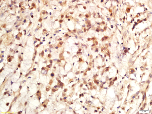
Figure 1. IHC analysis of Ki67/MKI67 using anti-Ki67/MKI67 antibody (A00254-1). Ki67/MKI67 was detected in a paraffin-embedded section of human colonic adenocarcinoma tissue. Heat mediated antigen retrieval was performed in EDTA buffer (pH 8.0, epitope retrieval solution). The tissue section was blocked with 10% goat serum. The tissue section was then incubated with 2 microg/ml rabbit anti-Ki67/MKI67 Antibody (A00254-1) overnight at 4°C. Peroxidase Conjugated Goat Anti-rabbit IgG was used as secondary antibody and incubated for 30 minutes at 37°C. The tissue section was developed using HRP Conjugated Rabbit IgG Super Vision Assay Kit (Catalog # SV0002) with DAB as the chromogen.
. Ki67/MKI67 was detected in a paraffin-embedded section of human lung cancer tissue. Heat mediated antigen retrieval was performed in EDTA buffer (pH 8.0, epitope retrieval solution). The tissue section was blocked with 10% goat serum. The tissue section was then incubated with 2 microg/ml rabbit anti-Ki67/MKI67 Antibody (A00254-1) overnight at 4°C. Peroxidase Conjugated Goat Anti-rabbit IgG was used as secondary antibody and incubated for 30 minutes at 37°C. The tissue section was developed using HRP Conjugated Rabbit IgG Super Vision Assay Kit (Catalog # SV0002) with DAB as the chromogen.")
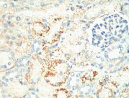
. Ki67/MKI67 was detected in a paraffin-embedded section of human renal carcinoma tissue. Heat mediated antigen retrieval was performed in EDTA buffer (pH 8.0, epitope retrieval solution). The tissue section was blocked with 10% goat serum. The tissue section was then incubated with 2 microg/ml rabbit anti-Ki67/MKI67 Antibody (A00254-1) overnight at 4°C. Peroxidase Conjugated Goat Anti-rabbit IgG was used as secondary antibody and incubated for 30 minutes at 37°C. The tissue section was developed using HRP Conjugated Rabbit IgG Super Vision Assay Kit (Catalog # SV0002) with DAB as the chromogen.")
. Ki67/MKI67 was detected in a paraffin-embedded section of human tonsil tissue. Heat mediated antigen retrieval was performed in EDTA buffer (pH 8.0, epitope retrieval solution). The tissue section was blocked with 10% goat serum. The tissue section was then incubated with 2 microg/ml rabbit anti-Ki67/MKI67 Antibody (A00254-1) overnight at 4°C. Peroxidase Conjugated Goat Anti-rabbit IgG was used as secondary antibody and incubated for 30 minutes at 37°C. The tissue section was developed using HRP Conjugated Rabbit IgG Super Vision Assay Kit (Catalog # SV0002) with DAB as the chromogen.")
. Ki67/MKI67 was detected in a paraffin-embedded section of human laryngeal squamous cell carcinoma tissue. Heat mediated antigen retrieval was performed in EDTA buffer (pH 8.0, epitope retrieval solution). The tissue section was blocked with 10% goat serum. The tissue section was then incubated with 2 microg/ml rabbit anti-Ki67/MKI67 Antibody (A00254-1) overnight at 4°C. Peroxidase Conjugated Goat Anti-rabbit IgG was used as secondary antibody and incubated for 30 minutes at 37°C. The tissue section was developed using HRP Conjugated Rabbit IgG Super Vision Assay Kit (Catalog # SV0002) with DAB as the chromogen.")
Figure 1. IHC analysis of Ki67/MKI67 using anti-Ki67/MKI67 antibody (A00254-1). Ki67/MKI67 was detected in a paraffin-embedded section of human colonic adenocarcinoma tissue. Heat mediated antigen retrieval was performed in EDTA buffer (pH 8.0, epitope retrieval solution). The tissue section was blocked with 10% goat serum. The tissue section was then incubated with 2 microg/ml rabbit anti-Ki67/MKI67 Antibody (A00254-1) overnight at 4°C. Peroxidase Conjugated Goat Anti-rabbit IgG was used as secondary antibody and incubated for 30 minutes at 37°C. The tissue section was developed using HRP Conjugated Rabbit IgG Super Vision Assay Kit (Catalog # SV0002) with DAB as the chromogen.
Anti-Ki67/MKI67 Antibody
A00254-1-CARRIER-FREE
ApplicationsELISA, ImmunoHistoChemistry
Product group Antibodies
ReactivityHuman
TargetMKI67
Overview
- SupplierBoster Bio
- Product NameAnti-Ki67/MKI67 Antibody
- Delivery Days Customer9
- Application Supplier NoteTested Species: In-house tested species with positive results. Other applications have not been tested. Optimal dilutions should be determined by end users.
- ApplicationsELISA, ImmunoHistoChemistry
- CertificationResearch Use Only
- ClonalityPolyclonal
- Concentration500 ug/ml
- Gene ID4288
- Target nameMKI67
- Target descriptionmarker of proliferation Ki-67
- Target synonymsKIA, MIB-, MIB-1, PPP1R105, proliferation marker protein Ki-67, Molecular Immunology Borstel antibody 1, antigen Ki67, antigen identified by monoclonal antibody Ki-67, proliferation-related Ki-67 antigen, protein phosphatase 1, regulatory subunit 105
- HostRabbit
- IsotypeIgG
- Protein IDP46013
- Protein NameProliferation marker protein Ki-67
- Scientific DescriptionBoster Bio Anti-Ki67/MKI67 Antibody Picoband® catalog # A00254-1. Tested in ELISA, IHC applications. This antibody reacts with Human.
- ReactivityHuman
- Storage Instruction-20°C,2°C to 8°C
- UNSPSC12352203
Related products
Product group Antibodies
Anti-Ki-67 Antibody118-10017
ApplicationsELISA, ImmunoHistoChemistry
ReactivityHuman
- SizePrice
Product group Antibodies
Anti-Ki67 AntibodyA104333
ApplicationsImmunoFluorescence, Western Blot, ImmunoCytoChemistry, ImmunoHistoChemistry
ReactivityHuman, Mouse, Rat
- SizePrice

Product group Antibodies
Anti-MKI67 AntibodyAMAB90870
ApplicationsImmunoCytoChemistry, ImmunoHistoChemistry
ReactivityHuman
TargetMKI67
- SizePrice
Product group Antibodies
References
Ki-67 Polyclonal AntibodyBS-2130R
ApplicationsImmunoFluorescence, ImmunoCytoChemistry, ImmunoHistoChemistry, ImmunoHistoChemistry Frozen, ImmunoHistoChemistry Paraffin
ReactivityHuman, Mouse, Rat
TargetMKI67
- SizePrice
Product group Antibodies
MKI67 Monoclonal AntibodyCSB-MA000222
ApplicationsELISA, ImmunoHistoChemistry
ReactivityHuman, Mouse, Rat
TargetMKI67
- SizePrice
Product group Antibodies
Mki67 Polyclonal AntibodyCAC07112
ApplicationsELISA, ImmunoHistoChemistry
TargetMKI67
- SizePrice
Product group Antibodies

ApplicationsImmunoHistoChemistry, ImmunoHistoChemistry Frozen, ImmunoHistoChemistry Paraffin
ReactivityHuman
TargetMKI67
- SizePrice
![IHC-P analysis of human invasive breast cancer of no special type (NST) tissue using GTX04361 Ki67 antibody [MSVA-267M] HistoMAX?. High Ki67 LI in a breast cancer of no special type (NST).](https://www.genetex.com/upload/website/prouct_img/normal/GTX04361/GTX04361_20230728_IHC-P_69_23072722_622.webp)
Product group Antibodies
ApplicationsImmunoHistoChemistry, ImmunoHistoChemistry Paraffin
ReactivityHuman
TargetMKI67
- SizePrice