

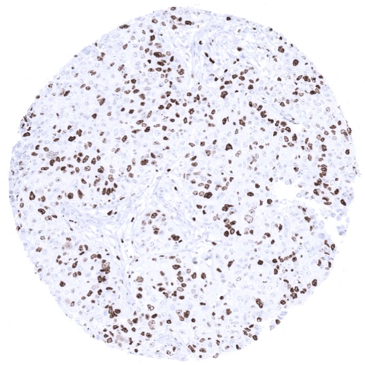
IHC-P analysis of human invasive breast cancer of no special type (NST) tissue using GTX04361 Ki67 antibody [MSVA-267M] HistoMAX?. High Ki67 LI in a breast cancer of no special type (NST).
![IHC-P analysis of human liver tissue using GTX04361 Ki67 antibody [MSVA-267M] HistoMAX?. The liver represents an ideal normal tissue control for Ki67 staining quality. Less than 1 of the hepatocytes should stain and cytoplasmatic staining should be absent.](https://www.genetex.com/upload/website/prouct_img/normal/GTX04361/GTX04361_20230728_IHC-P_199_23072723_261.webp "IHC-P analysis of human liver tissue using GTX04361 Ki67 antibody [MSVA-267M] HistoMAX?. The liver represents an ideal normal tissue control for Ki67 staining quality. Less than 1 of the hepatocytes should stain and cytoplasmatic staining should be absent.")
![IHC-P analysis of human tonsil tissue using GTX04361 Ki67 antibody [MSVA-267M] HistoMAX?. Ki67 stained cells are preferential seen in germinal centre and in supra basal cells of the surface epithelium.](https://www.genetex.com/upload/website/prouct_img/normal/GTX04361/GTX04361_20230728_IHC-P_329_23072723_477.webp "IHC-P analysis of human tonsil tissue using GTX04361 Ki67 antibody [MSVA-267M] HistoMAX?. Ki67 stained cells are preferential seen in germinal centre and in supra basal cells of the surface epithelium.")
IHC-P analysis of human invasive breast cancer of no special type (NST) tissue using GTX04361 Ki67 antibody [MSVA-267M] HistoMAX?. High Ki67 LI in a breast cancer of no special type (NST).
Ki67 antibody [MSVA-267M] HistoMAX(tm)
GTX04361
ApplicationsImmunoHistoChemistry, ImmunoHistoChemistry Paraffin
Product group Antibodies
ReactivityHuman
TargetMKI67
Overview
- SupplierGeneTex
- Product NameKi67 antibody [MSVA-267M] HistoMAX(tm)
- Delivery Days Customer9
- Application Supplier NoteIHC-P: 1:100-1:200. *Optimal dilutions/concentrations should be determined by the researcher.Not tested in other applications.
- ApplicationsImmunoHistoChemistry, ImmunoHistoChemistry Paraffin
- CertificationResearch Use Only
- ClonalityMonoclonal
- Clone IDMSVA-267M
- Concentration0.2 mg/ml
- ConjugateUnconjugated
- Gene ID4288
- Target nameMKI67
- Target descriptionmarker of proliferation Ki-67
- Target synonymsKIA, MIB-, MIB-1, PPP1R105, proliferation marker protein Ki-67, Molecular Immunology Borstel antibody 1, antigen Ki67, antigen identified by monoclonal antibody Ki-67, proliferation-related Ki-67 antigen, protein phosphatase 1, regulatory subunit 105
- HostMouse
- IsotypeIgG2b
- Protein IDP46013
- Protein NameProliferation marker protein Ki-67
- Scientific DescriptionThis gene encodes a nuclear protein that is associated with and may be necessary for cellular proliferation. Alternatively spliced transcript variants have been described. A related pseudogene exists on chromosome X. [provided by RefSeq, Mar 2009]
- ReactivityHuman
- Storage Instruction-20°C or -80°C,2°C to 8°C
- UNSPSC41116161
Datasheet
Related products
Product group Antibodies
Anti-Ki-67 Antibody118-10017
ApplicationsELISA, ImmunoHistoChemistry
ReactivityHuman
- SizePrice
Product group Antibodies
Anti-Ki67 AntibodyA104333
ApplicationsImmunoFluorescence, Western Blot, ImmunoCytoChemistry, ImmunoHistoChemistry
ReactivityHuman, Mouse, Rat
- SizePrice

Product group Antibodies
Anti-MKI67 AntibodyAMAB90870
ApplicationsImmunoCytoChemistry, ImmunoHistoChemistry
ReactivityHuman
TargetMKI67
- SizePrice

Product group Antibodies
Anti-Ki67/MKI67 AntibodyA00254-1-CARRIER-FREE
ApplicationsELISA, ImmunoHistoChemistry
ReactivityHuman
TargetMKI67
- SizePrice
Product group Antibodies
References
Ki-67 Polyclonal AntibodyBS-2130R
ApplicationsImmunoFluorescence, ImmunoCytoChemistry, ImmunoHistoChemistry, ImmunoHistoChemistry Frozen, ImmunoHistoChemistry Paraffin
ReactivityHuman, Mouse, Rat
TargetMKI67
- SizePrice
Product group Antibodies
MKI67 Monoclonal AntibodyCSB-MA000222
ApplicationsELISA, ImmunoHistoChemistry
ReactivityHuman, Mouse, Rat
TargetMKI67
- SizePrice
Product group Antibodies
Mki67 Polyclonal AntibodyCAC07112
ApplicationsELISA, ImmunoHistoChemistry
TargetMKI67
- SizePrice
![Ki67 antibody [SP6] detects Ki67 protein at nucleus in human cervical metaplasia (left) and carcinoma (right) by immunohistochemical analysis. Sample: Paraffin-embedded human cervix. Ki67 antibody [SP6] (GTX16667) diluted at 1:200.](https://www.genetex.com/upload/website/prouct_img/normal/GTX16667/GTX16667_20160307_IHC-P_w_23060620_422.webp)
Product group Antibodies
Ki67 antibody [SP6]GTX16667
ApplicationsFlow Cytometry, ImmunoFluorescence, Western Blot, ImmunoCytoChemistry, ImmunoHistoChemistry, ImmunoHistoChemistry Frozen, ImmunoHistoChemistry Paraffin
ReactivityChicken, Human, Mouse, Porcine, Rat
TargetMKI67
- SizePrice
![FACS analysis of HeLa cells using GTX17971 Ki67 antibody [MKI67/2462]. Blue : Primary antibody Red : Isotype control](https://www.genetex.com/upload/website/prouct_img/normal/GTX17971/GTX17971_20200115_FACS_189_w_23060620_847.webp)
Product group Antibodies
Ki67 antibody [MKI67/2462]GTX17971
ApplicationsFlow Cytometry, ImmunoFluorescence, Western Blot, ImmunoCytoChemistry, ImmunoHistoChemistry, ImmunoHistoChemistry Paraffin, Other Application
ReactivityHuman
TargetMKI67
- SizePrice