


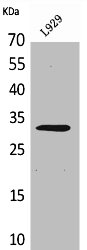
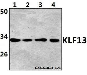
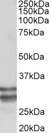
Figure 1. Western blot analysis of KLF13 using anti-KLF13 antibody (A07076-2). Electrophoresis was performed on a 5-20% SDS-PAGE gel at 70V (Stacking gel) / 90V (Resolving gel) for 2-3 hours. The sample well of each lane was loaded with 30 ug of sample under reducing conditions. Lane 1: human SH-SY5Y whole cell lysates, Lane 2: human Hela whole cell lysates, Lane 3: human HEL whole cell lysates, Lane 4: human HL-60 whole cell lysates, Lane 5: human PC-3 whole cell lysates, Lane 6: human Jurkat whole cell lysates, Lane 7: rat thymus tissue lysates, Lane 8: mouse thymus tissue lysates. After electrophoresis, proteins were transferred to a nitrocellulose membrane at 150 mA for 50-90 minutes. Blocked the membrane with 5% non-fat milk/TBS for 1.5 hour at RT. The membrane was incubated with rabbit anti-KLF13 antigen affinity purified polyclonal antibody (Catalog # A07076-2) at 0.5 microg/mL overnight at 4°C, then washed with TBS-0.1%Tween 3 times with 5 minutes each and probed with a goat anti-rabbit IgG-HRP secondary antibody at a dilution of 1:5000 for 1.5 hour at RT. The signal is developed using an Enhanced Chemiluminescent detection (ECL) kit (Catalog # EK1002) with Tanon 5200 system. A specific band was detected for KLF13 at approximately 38 kDa. The expected band size for KLF13 is at 31 kDa.
. KLF13 was detected in a paraffin-embedded section of rat thymus tissue. Heat mediated antigen retrieval was performed in EDTA buffer (pH 8.0, epitope retrieval solution). The tissue section was blocked with 10% goat serum. The tissue section was then incubated with 2 microg/ml rabbit anti-KLF13 Antibody (A07076-2) overnight at 4°C. Peroxidase Conjugated Goat Anti-rabbit IgG was used as secondary antibody and incubated for 30 minutes at 37°C. The tissue section was developed using HRP Conjugated Rabbit IgG Super Vision Assay Kit (Catalog # SV0002) with DAB as the chromogen.")
. Overlay histogram showing Hela cells stained with A07076-2 (Blue line). To facilitate intracellular staining, cells were fixed with 4% paraformaldehyde and permeabilized with permeabilization buffer. The cells were blocked with 10% normal goat serum. And then incubated with rabbit anti-KLF13 Antibody (A07076-2, 1 microg/1x106 cells) for 30 min at 20°C. DyLight®488 conjugated goat anti-rabbit IgG (BA1127, 5-10 microg/1x106 cells) was used as secondary antibody for 30 minutes at 20°C. Isotype control antibody (Green line) was rabbit IgG (1 microg/1x106) used under the same conditions. Unlabelled sample without incubation with primary antibody and secondary antibody (Red line) was used as a blank control.")
. Overlay histogram showing Neuro-2a cells stained with A07076-2 (Blue line). To facilitate intracellular staining, cells were fixed with 4% paraformaldehyde and permeabilized with permeabilization buffer. The cells were blocked with 10% normal goat serum. And then incubated with rabbit anti-KLF13 Antibody (A07076-2, 1 microg/1x106 cells) for 30 min at 20°C. DyLight®488 conjugated goat anti-rabbit IgG (BA1127, 5-10 microg/1x106 cells) was used as secondary antibody for 30 minutes at 20°C. Isotype control antibody (Green line) was rabbit IgG (1 microg/1x106) used under the same conditions. Unlabelled sample without incubation with primary antibody and secondary antibody (Red line) was used as a blank control.")
. Overlay histogram showing C6 cells stained with A07076-2 (Blue line). To facilitate intracellular staining, cells were fixed with 4% paraformaldehyde and permeabilized with permeabilization buffer. The cells were blocked with 10% normal goat serum. And then incubated with rabbit anti-KLF13 Antibody (A07076-2, 1 microg/1x106 cells) for 30 min at 20°C. DyLight®488 conjugated goat anti-rabbit IgG (BA1127, 5-10 microg/1x106 cells) was used as secondary antibody for 30 minutes at 20°C. Isotype control antibody (Green line) was rabbit IgG (1 microg/1x106) used under the same conditions. Unlabelled sample without incubation with primary antibody and secondary antibody (Red line) was used as a blank control.")
Figure 1. Western blot analysis of KLF13 using anti-KLF13 antibody (A07076-2). Electrophoresis was performed on a 5-20% SDS-PAGE gel at 70V (Stacking gel) / 90V (Resolving gel) for 2-3 hours. The sample well of each lane was loaded with 30 ug of sample under reducing conditions. Lane 1: human SH-SY5Y whole cell lysates, Lane 2: human Hela whole cell lysates, Lane 3: human HEL whole cell lysates, Lane 4: human HL-60 whole cell lysates, Lane 5: human PC-3 whole cell lysates, Lane 6: human Jurkat whole cell lysates, Lane 7: rat thymus tissue lysates, Lane 8: mouse thymus tissue lysates. After electrophoresis, proteins were transferred to a nitrocellulose membrane at 150 mA for 50-90 minutes. Blocked the membrane with 5% non-fat milk/TBS for 1.5 hour at RT. The membrane was incubated with rabbit anti-KLF13 antigen affinity purified polyclonal antibody (Catalog # A07076-2) at 0.5 microg/mL overnight at 4°C, then washed with TBS-0.1%Tween 3 times with 5 minutes each and probed with a goat anti-rabbit IgG-HRP secondary antibody at a dilution of 1:5000 for 1.5 hour at RT. The signal is developed using an Enhanced Chemiluminescent detection (ECL) kit (Catalog # EK1002) with Tanon 5200 system. A specific band was detected for KLF13 at approximately 38 kDa. The expected band size for KLF13 is at 31 kDa.
Anti-KLF13 Antibody Picoband(r)
A07076-2-CARRIER-FREE
ApplicationsFlow Cytometry, Western Blot, ELISA, ImmunoHistoChemistry
Product group Antibodies
ReactivityHuman, Mouse, Rat
TargetKLF13
Overview
- SupplierBoster Bio
- Product NameAnti-KLF13 Antibody Picoband(r)
- Delivery Days Customer9
- ApplicationsFlow Cytometry, Western Blot, ELISA, ImmunoHistoChemistry
- CertificationResearch Use Only
- ClonalityPolyclonal
- Concentration500 ug/ml
- Gene ID51621
- Target nameKLF13
- Target descriptionKLF transcription factor 13
- Target synonymsBTEB3, FKLF2, NSLP1, RFLAT-1, RFLAT1, Krueppel-like factor 13, BTE-binding protein 3, Kruppel like factor 13, RANTES factor of late activated T lymphocytes-1, basic transcription element binding protein 3, novel Sp1-like zinc finger transcription factor 1, transcription factor BTEB3, transcription factor NSLP1
- HostRabbit
- IsotypeIgG
- Protein IDQ9Y2Y9
- Protein NameKrueppel-like factor 13
- Scientific DescriptionBoster Bio Anti-KLF13 Antibody Picoband® catalog # A07076-2. Tested in ELISA, Flow Cytometry, IHC, WB applications. This antibody reacts with Human, Mouse, Rat. The brand Picoband indicates this is a premium antibody that guarantees superior quality, high affinity, and strong signals with minimal background in Western blot applications. Only our best-performing antibodies are designated as Picoband, ensuring unmatched performance.
- ReactivityHuman, Mouse, Rat
- Storage Instruction-20°C,2°C to 8°C
- UNSPSC12352203
Related products
Product group Antibodies
Anti-KLF13 Antibody101-10870
ApplicationsWestern Blot, ELISA
TargetKLF13
- SizePrice
Product group Antibodies
Klf13 Polyclonal AntibodyCAC11428
ApplicationsWestern Blot, ELISA, ImmunoHistoChemistry
TargetKLF13
- SizePrice
Product group Antibodies
Anti-RFLAT1 AntibodyBS-70812R
ApplicationsWestern Blot
ReactivityHuman, Mouse
TargetKLF13
- SizePrice
Product group Antibodies
KLF13 AntibodyCSB-PA005502
ApplicationsWestern Blot, ELISA
ReactivityHuman, Mouse
TargetKLF13
- SizePrice
Product group Antibodies
KLF13 (acetyl lys166) antibodyGTX66827
ApplicationsWestern Blot
ReactivityHuman, Mouse, Rat
TargetKLF13
- SizePrice
Product group Antibodies

KLF13 AntibodyLS-C673405
ApplicationsWestern Blot, ELISA, ImmunoHistoChemistry, ImmunoHistoChemistry Paraffin
ReactivityHuman
TargetKLF13
- SizePrice
Product group Antibodies
Anti-KLF13 AntibodyA85017
ApplicationsWestern Blot, ELISA
ReactivityHuman
- SizePrice