


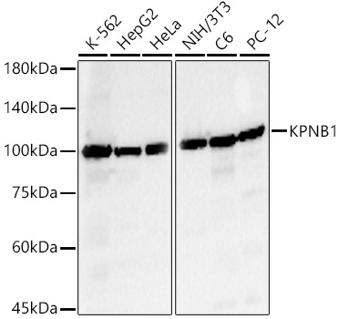
Figure 1. Western blot analysis of KPNB1 using anti-KPNB1 antibody (M01851-2). Electrophoresis was performed on a 5-20% SDS-PAGE gel at 70V (Stacking gel) / 90V (Resolving gel) for 2-3 hours. The sample well of each lane was loaded with 30 ug of sample under reducing conditions. Lane 1: human CACO-2 whole cell lysates, Lane 2: human K562 whole cell lysates, Lane 3: human A549 whole cell lysates, Lane 4: human A549 whole cell lysates, Lane 5: mouse testis tissue lysates, Lane 6: mouse lung tissue lysates. After electrophoresis, proteins were transferred to a nitrocellulose membrane at 150 mA for 50-90 minutes. Blocked the membrane with 5% non-fat milk/TBS for 1.5 hour at RT. The membrane was incubated with mouse anti-KPNB1 antigen affinity purified monoclonal antibody (Catalog # M01851-2) at 0.5 microg/mL overnight at 4°C, then washed with TBS-0.1%Tween 3 times with 5 minutes each and probed with a goat anti-mouse IgG-HRP secondary antibody at a dilution of 1:10000 for 1.5 hour at RT. The signal is developed using an Enhanced Chemiluminescent detection (ECL) kit (Catalog # EK1001) with Tanon 5200 system. A specific band was detected for KPNB1 at approximately 97 kDa. The expected band size for KPNB1 is at 97 kDa.
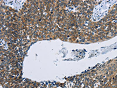
. KPNB1 was detected in a paraffin-embedded section of human thyroiditis tissue. Heat mediated antigen retrieval was performed in EDTA buffer (pH 8.0, epitope retrieval solution). The tissue section was blocked with 10% goat serum. The tissue section was then incubated with 2 microg/ml mouse anti-KPNB1 Antibody (M01851-2) overnight at 4°C. Peroxidase Conjugated Goat Anti-mouse IgG was used as secondary antibody and incubated for 30 minutes at 37°C. The tissue section was developed using HRP Conjugated Mouse IgG Super Vision Assay Kit (Catalog # SV0001) with DAB as the chromogen.")
. KPNB1 was detected in a paraffin-embedded section of human squamous cell lung carcinoma tissue. Heat mediated antigen retrieval was performed in EDTA buffer (pH 8.0, epitope retrieval solution). The tissue section was blocked with 10% goat serum. The tissue section was then incubated with 2 microg/ml mouse anti-KPNB1 Antibody (M01851-2) overnight at 4°C. Peroxidase Conjugated Goat Anti-mouse IgG was used as secondary antibody and incubated for 30 minutes at 37°C. The tissue section was developed using HRP Conjugated Mouse IgG Super Vision Assay Kit (Catalog # SV0001) with DAB as the chromogen.")
. KPNB1 was detected in a paraffin-embedded section of human renal carcinoma tissue. Heat mediated antigen retrieval was performed in EDTA buffer (pH 8.0, epitope retrieval solution). The tissue section was blocked with 10% goat serum. The tissue section was then incubated with 2 microg/ml mouse anti-KPNB1 Antibody (M01851-2) overnight at 4°C. Peroxidase Conjugated Goat Anti-mouse IgG was used as secondary antibody and incubated for 30 minutes at 37°C. The tissue section was developed using HRP Conjugated Mouse IgG Super Vision Assay Kit (Catalog # SV0001) with DAB as the chromogen.")
. KPNB1 was detected in a paraffin-embedded section of human breast cancer tissue. Heat mediated antigen retrieval was performed in EDTA buffer (pH 8.0, epitope retrieval solution). The tissue section was blocked with 10% goat serum. The tissue section was then incubated with 2 microg/ml mouse anti-KPNB1 Antibody (M01851-2) overnight at 4°C. Peroxidase Conjugated Goat Anti-mouse IgG was used as secondary antibody and incubated for 30 minutes at 37°C. The tissue section was developed using HRP Conjugated Mouse IgG Super Vision Assay Kit (Catalog # SV0001) with DAB as the chromogen.")
. KPNB1 was detected in a paraffin-embedded section of human colorectal adenocarcinoma tissue. Heat mediated antigen retrieval was performed in EDTA buffer (pH 8.0, epitope retrieval solution). The tissue section was blocked with 10% goat serum. The tissue section was then incubated with 2 microg/ml mouse anti-KPNB1 Antibody (M01851-2) overnight at 4°C. Peroxidase Conjugated Goat Anti-mouse IgG was used as secondary antibody and incubated for 30 minutes at 37°C. The tissue section was developed using HRP Conjugated Mouse IgG Super Vision Assay Kit (Catalog # SV0001) with DAB as the chromogen.")
. KPNB1 was detected in a paraffin-embedded section of human laryngeal squamous cell carcinoma tissue. Heat mediated antigen retrieval was performed in EDTA buffer (pH 8.0, epitope retrieval solution). The tissue section was blocked with 10% goat serum. The tissue section was then incubated with 2 microg/ml mouse anti-KPNB1 Antibody (M01851-2) overnight at 4°C. Peroxidase Conjugated Goat Anti-mouse IgG was used as secondary antibody and incubated for 30 minutes at 37°C. The tissue section was developed using HRP Conjugated Mouse IgG Super Vision Assay Kit (Catalog # SV0001) with DAB as the chromogen.")
. KPNB1 was detected in a paraffin-embedded section of human ovary tissue. Heat mediated antigen retrieval was performed in EDTA buffer (pH 8.0, epitope retrieval solution). The tissue section was blocked with 10% goat serum. The tissue section was then incubated with 2 microg/ml mouse anti-KPNB1 Antibody (M01851-2) overnight at 4°C. Peroxidase Conjugated Goat Anti-mouse IgG was used as secondary antibody and incubated for 30 minutes at 37°C. The tissue section was developed using HRP Conjugated Mouse IgG Super Vision Assay Kit (Catalog # SV0001) with DAB as the chromogen.")
. KPNB1 was detected in a paraffin-embedded section of human placenta tissue. Heat mediated antigen retrieval was performed in EDTA buffer (pH 8.0, epitope retrieval solution). The tissue section was blocked with 10% goat serum. The tissue section was then incubated with 2 microg/ml mouse anti-KPNB1 Antibody (M01851-2) overnight at 4°C. Peroxidase Conjugated Goat Anti-mouse IgG was used as secondary antibody and incubated for 30 minutes at 37°C. The tissue section was developed using HRP Conjugated Mouse IgG Super Vision Assay Kit (Catalog # SV0001) with DAB as the chromogen.")
. KPNB1 was detected in a paraffin-embedded section of human spleen tissue. Heat mediated antigen retrieval was performed in EDTA buffer (pH 8.0, epitope retrieval solution). The tissue section was blocked with 10% goat serum. The tissue section was then incubated with 2 microg/ml mouse anti-KPNB1 Antibody (M01851-2) overnight at 4°C. Peroxidase Conjugated Goat Anti-mouse IgG was used as secondary antibody and incubated for 30 minutes at 37°C. The tissue section was developed using HRP Conjugated Mouse IgG Super Vision Assay Kit (Catalog # SV0001) with DAB as the chromogen.")
Figure 1. Western blot analysis of KPNB1 using anti-KPNB1 antibody (M01851-2). Electrophoresis was performed on a 5-20% SDS-PAGE gel at 70V (Stacking gel) / 90V (Resolving gel) for 2-3 hours. The sample well of each lane was loaded with 30 ug of sample under reducing conditions. Lane 1: human CACO-2 whole cell lysates, Lane 2: human K562 whole cell lysates, Lane 3: human A549 whole cell lysates, Lane 4: human A549 whole cell lysates, Lane 5: mouse testis tissue lysates, Lane 6: mouse lung tissue lysates. After electrophoresis, proteins were transferred to a nitrocellulose membrane at 150 mA for 50-90 minutes. Blocked the membrane with 5% non-fat milk/TBS for 1.5 hour at RT. The membrane was incubated with mouse anti-KPNB1 antigen affinity purified monoclonal antibody (Catalog # M01851-2) at 0.5 microg/mL overnight at 4°C, then washed with TBS-0.1%Tween 3 times with 5 minutes each and probed with a goat anti-mouse IgG-HRP secondary antibody at a dilution of 1:10000 for 1.5 hour at RT. The signal is developed using an Enhanced Chemiluminescent detection (ECL) kit (Catalog # EK1001) with Tanon 5200 system. A specific band was detected for KPNB1 at approximately 97 kDa. The expected band size for KPNB1 is at 97 kDa.
Anti-KPNB1 Antibody Picoband(r) (monoclonal, 3I11F2)
M01851-2-DYLIGHT550
ApplicationsFlow Cytometry, ImmunoFluorescence, Western Blot, ImmunoCytoChemistry, ImmunoHistoChemistry
Product group Antibodies
ReactivityHuman, Mouse, Rat
TargetKPNB1
Overview
- SupplierBoster Bio
- Product NameAnti-KPNB1 Antibody Picoband(r) (monoclonal, 3I11F2)
- Delivery Days Customer9
- ApplicationsFlow Cytometry, ImmunoFluorescence, Western Blot, ImmunoCytoChemistry, ImmunoHistoChemistry
- CertificationResearch Use Only
- ClonalityMonoclonal
- Clone ID3I11F2
- Concentration500 ug/ml
- ConjugateDyLight 550
- Gene ID3837
- Target nameKPNB1
- Target descriptionkaryopherin subunit beta 1
- Target synonymsIMB1, IPO1, IPOB, Impnb, NTF97, importin subunit beta-1, PTAC97, importin 1, importin 90, importin beta-1 subunit, karyopherin (importin) beta 1, nuclear factor p97, pore targeting complex 97 kDa subunit
- HostMouse
- IsotypeIgG2b
- Protein IDQ14974
- Protein NameImportin subunit beta-1
- Scientific DescriptionBoster Bio Anti-KPNB1 Antibody Picoband® (monoclonal, 3I11F2) catalog # M01851-2. Tested in Flow Cytometry, IF, IHC, ICC, WB applications. This antibody reacts with Human, Mouse, Rat. The brand Picoband indicates this is a premium antibody that guarantees superior quality, high affinity, and strong signals with minimal background in Western blot applications. Only our best-performing antibodies are designated as Picoband, ensuring unmatched performance.
- ReactivityHuman, Mouse, Rat
- Storage Instruction-20°C,2°C to 8°C
- UNSPSC12352203
Related products
Product group Antibodies
Anti-KPNB1 Antibody144-08610
ApplicationsWestern Blot, ImmunoHistoChemistry
ReactivityHuman, Mouse, Rat
TargetKPNB1
- SizePrice
Product group Antibodies
KPNB1 Polyclonal AntibodyCAC15768
ApplicationsWestern Blot, ELISA, ImmunoHistoChemistry
TargetKPNB1
- SizePrice
Product group Antibodies

ApplicationsFlow Cytometry, ImmunoFluorescence, Western Blot, ELISA, ImmunoHistoChemistry
ReactivityBovine, Canine, Human, Mouse, Rat
TargetKPNB1
- SizePrice
Product group Antibodies
References
Importin beta 1 antibodyGTX133733
ApplicationsWestern Blot, ImmunoHistoChemistry, ImmunoHistoChemistry Paraffin
ReactivityHuman, Mouse
TargetKPNB1
- SizePrice
Product group Antibodies
Anti-KPNB1 AntibodyA16248
ApplicationsImmunoFluorescence, Western Blot, ImmunoCytoChemistry, ImmunoHistoChemistry
ReactivityHuman, Mouse, Rat
- SizePrice

Product group Antibodies
Anti-KPNB1 Antibody Picoband(r)A01851-2-CARRIER-FREE
ApplicationsFlow Cytometry, ImmunoFluorescence, Western Blot, ELISA, ImmunoCytoChemistry
ReactivityHuman, Mouse, Rat
TargetKPNB1
- SizePrice
Product group Antibodies
KPNB1 AntibodyCSB-PA244093
ApplicationsELISA, ImmunoHistoChemistry
ReactivityHuman, Mouse, Rat
TargetKPNB1
- SizePrice
Product group Antibodies
NTF97 Polyclonal AntibodyBS-3695R
ApplicationsImmunoFluorescence, Western Blot, ELISA, ImmunoCytoChemistry, ImmunoHistoChemistry, ImmunoHistoChemistry Frozen, ImmunoHistoChemistry Paraffin
ReactivityBovine, Chicken, Equine, Human, Mouse, Porcine, Rat, Sheep
TargetKPNB1
- SizePrice

Product group Antibodies
Anti-KPNB1 AntibodyHPA050302
ApplicationsImmunoCytoChemistry
ReactivityHuman
TargetKPNB1
- SizePrice