


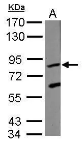
Figure 1. Western blot analysis of KRIT1 using anti-KRIT1 antibody (PB10074). Electrophoresis was performed on a 5-20% SDS-PAGE gel at 70V (Stacking gel) / 90V (Resolving gel) for 2-3 hours. The sample well of each lane was loaded with 30 ug of sample under reducing conditions. Lane 1: rat cardiac muscle tissue lysates, Lane 2: NIH3T3 whole cell lysates, Lane 3: HELA whole cell lysates. After electrophoresis, proteins were transferred to a nitrocellulose membrane at 150 mA for 50-90 minutes. Blocked the membrane with 5% non-fat milk/TBS for 1.5 hour at RT. The membrane was incubated with rabbit anti-KRIT1 antigen affinity purified polyclonal antibody (Catalog # PB10074) at 0.5 microg/mL overnight at 4°C, then washed with TBS-0.1%Tween 3 times with 5 minutes each and probed with a goat anti-rabbit IgG-HRP secondary antibody at a dilution of 1:5000 for 1.5 hour at RT. The signal is developed using an Enhanced Chemiluminescent detection (ECL) kit (Catalog # EK1002) with Tanon 5200 system. A specific band was detected for KRIT1 at approximately 84 kDa. The expected band size for KRIT1 is at 84 kDa.
Figure 1. Western blot analysis of KRIT1 using anti-KRIT1 antibody (PB10074). Electrophoresis was performed on a 5-20% SDS-PAGE gel at 70V (Stacking gel) / 90V (Resolving gel) for 2-3 hours. The sample well of each lane was loaded with 30 ug of sample under reducing conditions. Lane 1: rat cardiac muscle tissue lysates, Lane 2: NIH3T3 whole cell lysates, Lane 3: HELA whole cell lysates. After electrophoresis, proteins were transferred to a nitrocellulose membrane at 150 mA for 50-90 minutes. Blocked the membrane with 5% non-fat milk/TBS for 1.5 hour at RT. The membrane was incubated with rabbit anti-KRIT1 antigen affinity purified polyclonal antibody (Catalog # PB10074) at 0.5 microg/mL overnight at 4°C, then washed with TBS-0.1%Tween 3 times with 5 minutes each and probed with a goat anti-rabbit IgG-HRP secondary antibody at a dilution of 1:5000 for 1.5 hour at RT. The signal is developed using an Enhanced Chemiluminescent detection (ECL) kit (Catalog # EK1002) with Tanon 5200 system. A specific band was detected for KRIT1 at approximately 84 kDa. The expected band size for KRIT1 is at 84 kDa.
Anti-KRIT1 Antibody Picoband(r)
PB10074-CARRIER-FREE
ApplicationsWestern Blot
Product group Antibodies
ReactivityHamster, Human, Mouse, Rat
TargetKRIT1
Overview
- SupplierBoster Bio
- Product NameAnti-KRIT1 Antibody Picoband(r)
- Delivery Days Customer9
- Application Supplier NoteTested Species: In-house tested species with positive results. Other applications have not been tested. Optimal dilutions should be determined by end users.
- ApplicationsWestern Blot
- CertificationResearch Use Only
- ClonalityPolyclonal
- Concentration500 ug/ml
- Gene ID889
- Target nameKRIT1
- Target descriptionKRIT1 ankyrin repeat containing
- Target synonymsCAM, CCM1, krev interaction trapped protein 1, ankyrin repeat-containing protein Krit1, cerebral cavernous malformations 1 protein, krev interaction trapped 1
- HostRabbit
- IsotypeIgG
- Protein IDO00522
- Protein NameKrev interaction trapped protein 1
- Scientific DescriptionBoster Bio Anti-KRIT1 Antibody Picoband® catalog # PB10074. Tested in WB applications. This antibody reacts with Human, Mouse, Rat. The brand Picoband indicates this is a premium antibody that guarantees superior quality, high affinity, and strong signals with minimal background in Western blot applications. Only our best-performing antibodies are designated as Picoband, ensuring unmatched performance.
- ReactivityHamster, Human, Mouse, Rat
- Storage Instruction-20°C,2°C to 8°C
- UNSPSC12352203
Related products
Product group Antibodies
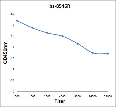
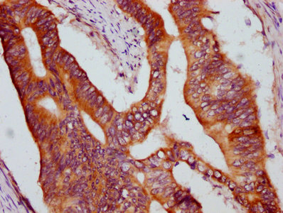
KRIT1 Polyclonal AntibodyBS-8546R
ApplicationsImmunoFluorescence, Western Blot, ELISA, ImmunoCytoChemistry, ImmunoHistoChemistry, ImmunoHistoChemistry Frozen, ImmunoHistoChemistry Paraffin
ReactivityBovine, Canine, Chicken, Equine, Human, Mouse, Porcine, Rabbit, Rat
TargetKRIT1
- SizePrice
Product group Antibodies
KRIT1 AntibodyCSB-PA012501LA01HU
ApplicationsImmunoFluorescence, ELISA, ImmunoHistoChemistry
ReactivityHuman
TargetKRIT1
- SizePrice

Product group Antibodies
Anti-KRIT1 AntibodyHPA049606
ApplicationsImmunoHistoChemistry
ReactivityHuman
TargetKRIT1
- SizePrice
Product group Antibodies

CCM1 / KRIT1 AntibodyLS-C346127
ApplicationsImmunoFluorescence, Western Blot
ReactivityHuman, Mouse, Rat
TargetKRIT1
- SizePrice
Product group Antibodies
References
KRIT1 antibody [C1C3]GTX109560
ApplicationsElectron Microscopy, Western Blot
ReactivityHuman
TargetKRIT1
- SizePrice