


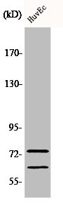
Figure 1. Western blot analysis of Lamin A/C using anti-Lamin A/C antibody (PB9280). Electrophoresis was performed on a 5-20% SDS-PAGE gel at 70V (Stacking gel) / 90V (Resolving gel) for 2-3 hours. The sample well of each lane was loaded with 30 ug of sample under reducing conditions. Lane 1: human placenta tissue lysates, Lane 2: human PC-3 whole cell lysates, Lane 3: human Hela whole cell lysates, Lane 4: human HepG2 whole cell lysates. After electrophoresis, proteins were transferred to a nitrocellulose membrane at 150 mA for 50-90 minutes. Blocked the membrane with 5% non-fat milk/TBS for 1.5 hour at RT. The membrane was incubated with rabbit anti-Lamin A/C antigen affinity purified polyclonal antibody (Catalog # PB9280) at 0.5 microg/mL overnight at 4°C, then washed with TBS-0.1%Tween 3 times with 5 minutes each and probed with a goat anti-rabbit IgG-HRP secondary antibody at a dilution of 1:5000 for 1.5 hour at RT. The signal is developed using an Enhanced Chemiluminescent detection (ECL) kit (Catalog # EK1002) with Tanon 5200 system. A specific band was detected for Lamin A/C at approximately 74 kDa, 63 kDa. The expected band size for Lamin A/C is at 74 kDa.
. Lamin A/C was detected in a paraffin-embedded section of Mouse Intestine tissue. Heat mediated antigen retrieval was performed in EDTA buffer (pH 8.0, epitope retrieval solution). The tissue section was blocked with 10% goat serum. The tissue section was then incubated with 1 microg/ml rabbit anti-Lamin A/C Antibody (PB9280) overnight at 4°C. Peroxidase Conjugated Goat Anti-rabbit IgG was used as secondary antibody and incubated for 30 minutes at 37°C. The tissue section was developed using HRP Conjugated Rabbit IgG Super Vision Assay Kit (Catalog # SV0002) with DAB as the chromogen.")
. Lamin A/C was detected in a paraffin-embedded section of Rat Intestine tissue. Heat mediated antigen retrieval was performed in EDTA buffer (pH 8.0, epitope retrieval solution). The tissue section was blocked with 10% goat serum. The tissue section was then incubated with 1 microg/ml rabbit anti-Lamin A/C Antibody (PB9280) overnight at 4°C. Peroxidase Conjugated Goat Anti-rabbit IgG was used as secondary antibody and incubated for 30 minutes at 37°C. The tissue section was developed using HRP Conjugated Rabbit IgG Super Vision Assay Kit (Catalog # SV0002) with DAB as the chromogen.")
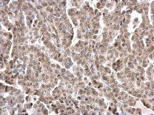
. Lamin A/C was detected in a paraffin-embedded section of Human Mammary Cancer tissue. Heat mediated antigen retrieval was performed in EDTA buffer (pH 8.0, epitope retrieval solution). The tissue section was blocked with 10% goat serum. The tissue section was then incubated with 1 microg/ml rabbit anti-Lamin A/C Antibody (PB9280) overnight at 4°C. Peroxidase Conjugated Goat Anti-rabbit IgG was used as secondary antibody and incubated for 30 minutes at 37°C. The tissue section was developed using HRP Conjugated Rabbit IgG Super Vision Assay Kit (Catalog # SV0002) with DAB as the chromogen.")
. Lamin A/C was detected in immunocytochemical section of A431 cells. Enzyme antigen retrieval was performed using IHC enzyme antigen retrieval reagent (AR0022) for 15 mins. The cells were blocked with 10% goat serum. And then incubated with 2microg/mL rabbit anti-Lamin A/C Antibody (PB9280) overnight at 4°C. DyLight?488 Conjugated Goat Anti-Rabbit IgG (BA1127) was used as secondary antibody at 1:100 dilution and incubated for 30 minutes at 37°C. The section was counterstained with DAPI. Visualize using a fluorescence microscope and filter sets appropriate for the label used.")
. Lamin A/C was detected in immunocytochemical section of NIH3T3 cells. Enzyme antigen retrieval was performed using IHC enzyme antigen retrieval reagent (AR0022) for 15 mins. The cells were blocked with 10% goat serum. And then incubated with 2microg/mL rabbit anti-Lamin A/C Antibody (PB9280) overnight at 4°C. DyLight?594 Conjugated Goat Anti-Rabbit IgG (BA1142) was used as secondary antibody at 1:100 dilution and incubated for 30 minutes at 37°C. The section was counterstained with DAPI. Visualize using a fluorescence microscope and filter sets appropriate for the label used.")
. Overlay histogram showing THP-1 cells stained with PB9280 (Blue line).The cells were blocked with 10% normal goat serum. And then incubated with rabbit anti-Lamin A/C Antibody (PB9280,1microg/1x106 cells) for 30 min at 20°C. DyLight?488 conjugated goat anti-rabbit IgG (BA1127, 5-10microg/1x106 cells) was used as secondary antibody for 30 minutes at 20°C. Isotype control antibody (Green line) was rabbit IgG (1microg/1x106) used under the same conditions. Unlabelled sample (Red line) was also used as a control.")
Figure 1. Western blot analysis of Lamin A/C using anti-Lamin A/C antibody (PB9280). Electrophoresis was performed on a 5-20% SDS-PAGE gel at 70V (Stacking gel) / 90V (Resolving gel) for 2-3 hours. The sample well of each lane was loaded with 30 ug of sample under reducing conditions. Lane 1: human placenta tissue lysates, Lane 2: human PC-3 whole cell lysates, Lane 3: human Hela whole cell lysates, Lane 4: human HepG2 whole cell lysates. After electrophoresis, proteins were transferred to a nitrocellulose membrane at 150 mA for 50-90 minutes. Blocked the membrane with 5% non-fat milk/TBS for 1.5 hour at RT. The membrane was incubated with rabbit anti-Lamin A/C antigen affinity purified polyclonal antibody (Catalog # PB9280) at 0.5 microg/mL overnight at 4°C, then washed with TBS-0.1%Tween 3 times with 5 minutes each and probed with a goat anti-rabbit IgG-HRP secondary antibody at a dilution of 1:5000 for 1.5 hour at RT. The signal is developed using an Enhanced Chemiluminescent detection (ECL) kit (Catalog # EK1002) with Tanon 5200 system. A specific band was detected for Lamin A/C at approximately 74 kDa, 63 kDa. The expected band size for Lamin A/C is at 74 kDa.
Anti-Lamin A+C/LMNA Antibody Picoband(r)
PB9280-CARRIER-FREE
ApplicationsFlow Cytometry, ImmunoFluorescence, Western Blot, ImmunoCytoChemistry, ImmunoHistoChemistry
Product group Antibodies
ReactivityHuman, Mouse, Rat
TargetLMNA
Overview
- SupplierBoster Bio
- Product NameAnti-Lamin A+C/LMNA Antibody Picoband(r)
- Delivery Days Customer9
- Application Supplier NoteWB: The detection limit for Lamin A/C is approximately 0.1ng/lane under reducing conditions. Tested Species: In-house tested species with positive results. By Heat: Boiling the paraffin sections in 10mM citrate buffer, pH6.0, for 20mins is required for the staining of formalin/paraffin sections. Other applications have not been tested. Optimal dilutions should be determined by end users.
- ApplicationsFlow Cytometry, ImmunoFluorescence, Western Blot, ImmunoCytoChemistry, ImmunoHistoChemistry
- CertificationResearch Use Only
- ClonalityPolyclonal
- Concentration500 ug/ml
- Gene ID4000
- Target nameLMNA
- Target descriptionlamin A/C
- Target synonymsCDCD1, CDDC, CMD1A, CMT2B1, EMD2, FPL, FPLD, FPLD2, HGPS, IDC, LDP1, LFP, LGMD1B, LMN1, LMNC, LMNL1, MADA, PRO1, lamin, 70 kDa lamin, epididymis secretory sperm binding protein, lamin A/C-like 1, lamin C, mandibuloacral dysplasia type A, prelamin-A/C, progerin, renal carcinoma antigen NY-REN-32
- HostRabbit
- IsotypeIgG
- Protein IDP02545
- Protein NamePrelamin-A/C
- Scientific DescriptionBoster Bio Anti-Lamin A+C/LMNA Antibody Picoband® catalog # PB9280. Tested in Flow Cytometry, IF, IHC, ICC, WB applications. This antibody reacts with Human, Mouse, Rat. The brand Picoband indicates this is a premium antibody that guarantees superior quality, high affinity, and strong signals with minimal background in Western blot applications. Only our best-performing antibodies are designated as Picoband, ensuring unmatched performance.
- ReactivityHuman, Mouse, Rat
- Storage Instruction-20°C,2°C to 8°C
- UNSPSC12352203
Related products
Product group Antibodies
LMNA AntibodyCSB-PA003130
ApplicationsImmunoFluorescence, Western Blot, ELISA, ImmunoHistoChemistry
ReactivityHuman, Mouse, Rat
TargetLMNA
- SizePrice
Product group Antibodies
Anti-Lamin A AntibodyA96570
ApplicationsWestern Blot, ELISA
ReactivityHuman, Mouse, Rat
- SizePrice
Product group Antibodies

Anti-Lamin A [133A2]AB03358-3.0
ApplicationsFlow Cytometry, ImmunoFluorescence, Western Blot, ImmunoHistoChemistry
ReactivityBovine, Canine, Human, Mouse, Rat
TargetLMNA
- SizePrice
Product group Antibodies

LMNA / Lamin A+C AntibodyLS-C767909
ApplicationsWestern Blot
ReactivityHuman
TargetLMNA
- SizePrice
Product group Antibodies

ApplicationsELISA, ImmunoHistoChemistry
ReactivityHuman, Mouse, Rat
TargetLMNA
- SizePrice

Product group Antibodies
Anti-LMNA AntibodyHPA006660
ApplicationsWestern Blot, ImmunoHistoChemistry
ReactivityHuman
TargetLMNA
- SizePrice
Product group Antibodies

Mouse anti Lamin AMUB1101P-CE/IVD
ApplicationsFlow Cytometry, Western Blot, ImmunoCytoChemistry, ImmunoHistoChemistry, ImmunoHistoChemistry Frozen, ImmunoHistoChemistry Paraffin
ReactivityBovine, Canine, Human, Mouse, Rat
TargetLMNA
- SizePrice
Product group Antibodies
LMNA Polyclonal AntibodyCAC15780
ApplicationsImmunoFluorescence, Western Blot, ELISA, ImmunoHistoChemistry
ReactivityRat
TargetLMNA
- SizePrice
Product group Antibodies
Lamin A + C antibodyGTX101126
ApplicationsImmunoFluorescence, ImmunoPrecipitation, Western Blot, ImmunoCytoChemistry, ImmunoHistoChemistry, ImmunoHistoChemistry Paraffin
ReactivityHuman, Mouse
TargetLMNA
- SizePrice